
|
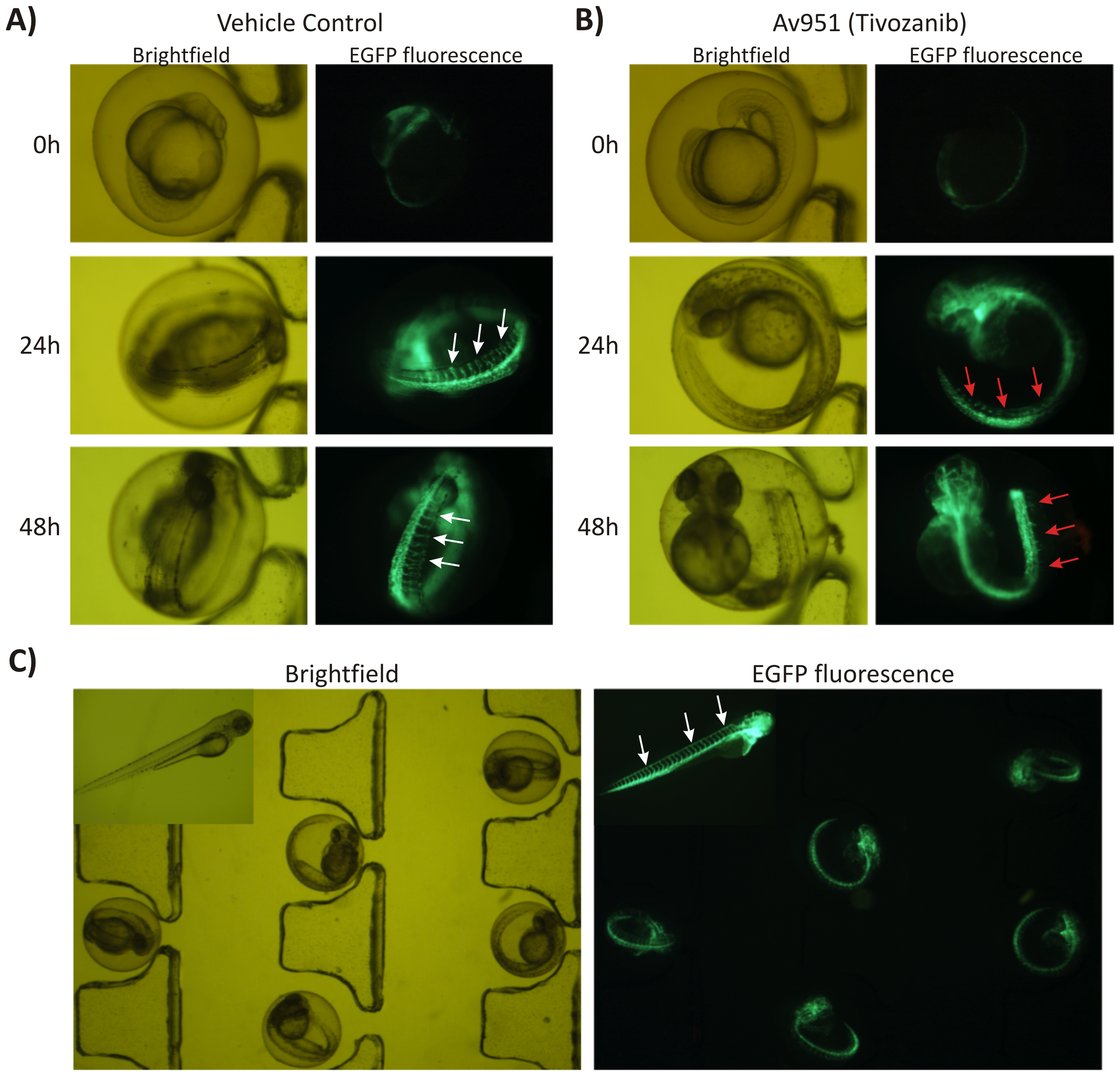
Fig. 6
On-chip angiogenesis assay using transgenic zebrafish line:
A) The transgenic fli1a:EGFP embryos at 16 hpf were loaded, immobilized and continuously perfused on a chip with E3 media containing vehicle control (DMSO); B) The transgenic fli1a:EGFP embryos at 16 hpf were loaded, immobilized and continuously perfused on a chip with a 1 μM of selective VEGFR inhibitor AV951 (Tivozanib, AVEO Pharmaceuticals Inc). Fluorescent and brightfield images were acquired at 0, 24 and 48 hours intervals. The optical transparency of embryos coupled with hydrodynamic immobilization on a chip array allowed for convenient microscopic visualization of characteristic patterns of intersegmental vessels (ISV, white arrows) and their AV951-induced inhibition (red arrows); C) Fli1a:EGFP transgenic embryos arrayed and hydrodynamically immobilized on a chip-based device. Developing patterns of intersegmental vessels are clearly visible even at the low magnification. Inset shows high magnification of hatched fli1a:EGFP larva with fully developed pattern of vasculature (white arrows).