
|
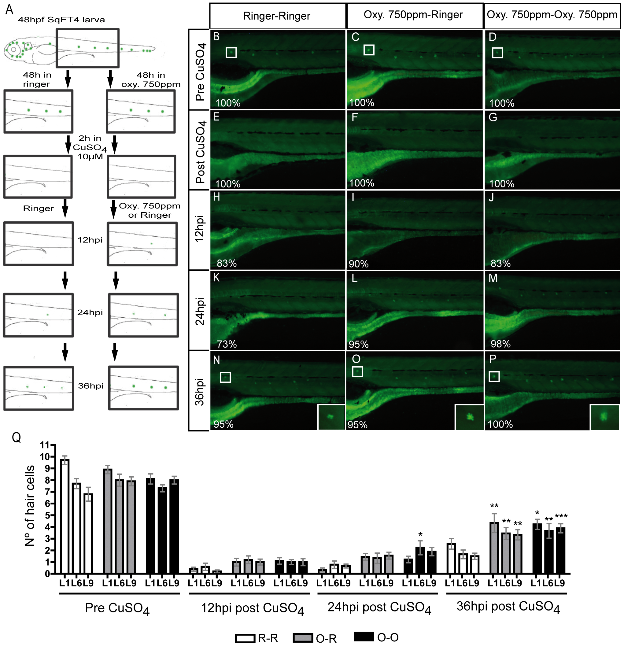
Fig. 5
Oxytetracycline induced inflammation increase regeneration capacity.
(A) Scheme of the regeneration assay developed. Incubation of SqET4 transgenic larvae (that express GFP in neuromast hair cells, HC) in oxytetracycline 750 ppm increases the rate of GFP/hair cells appearance. (B–D) HC before copper treatment. (E–G) CuSO4 treatment eliminates all GFP positive cells. (H–M, Q) Although we observe a tendency, there is no significant difference in the number of regenerated HC. (N–Q) At 36hpi the regeneration capacity of HC is significantly increased on oxytetracycline exposed larvae. (Q) The quantification of hair cells was performed in three neuromast; L1, L6 and L9. White box indicate L1 neuromast hair cells. The numbers of larvae that presented the phenotype shown is expressed as a percentage. * p<0.05, ** p<0.01, ***p<0.001.