
|
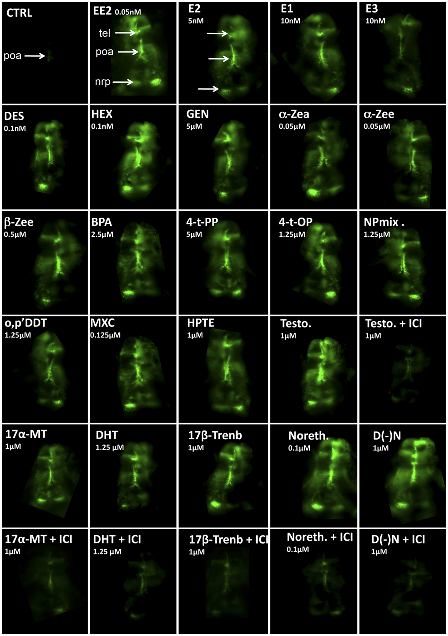
Fig. 2
In vivo imaging of 5-dpf old live transgenic cyp19a1b-GFP zebrafish embryos exposed to chemicals inducing GFP expression in radial glial progenitors.
Dorsal views (anterior to the top) of the telencephalon (tel), preoptic area (poa), and nucleus recessus posterioris (nrp) of the caudal hypothalamus. For each chemical the concentration used is indicated. CTRL: solvent control, EE2: 17α-ethinylestradiol, E2: 17β-estradiol, E1: estrone, E3: estriol, DES: diethystilbestrol, HEX: hexestrol, GEN: genistein, α-ZEA: α-zearalenol, α-ZEE: α-zearalanol, β-ZEE: β-zearalanol, BPA: bisphenol A, 4-t-PP: 4-t-pentylphenol, 4-t-OP, 4-t octylphenol, NPmix: mixture of nonylphenol, o,p′DDT: 1,1,1-Trichloro-2-(2-chlorophenyl)-2-(4- chlorophenyl)ethane,MXC: methoxychlor, HPTE 2,2-bis(p-hydroxyphenyl)-1,1,1-trichloro ethane,Testo: testosterone, DHT: dihydotestosterone, 17α-MT: 17α-methyltestosterone, 17β-Trenb: 17β-trenbolone, Noreth.: 17α-Ethynyl-19-nortestosterone, D(-)N: 13β-Ethyl-17α-ethynyl-17β-hydroxygon-4-e n-3-one,ICI (ICI 182-780).