
|
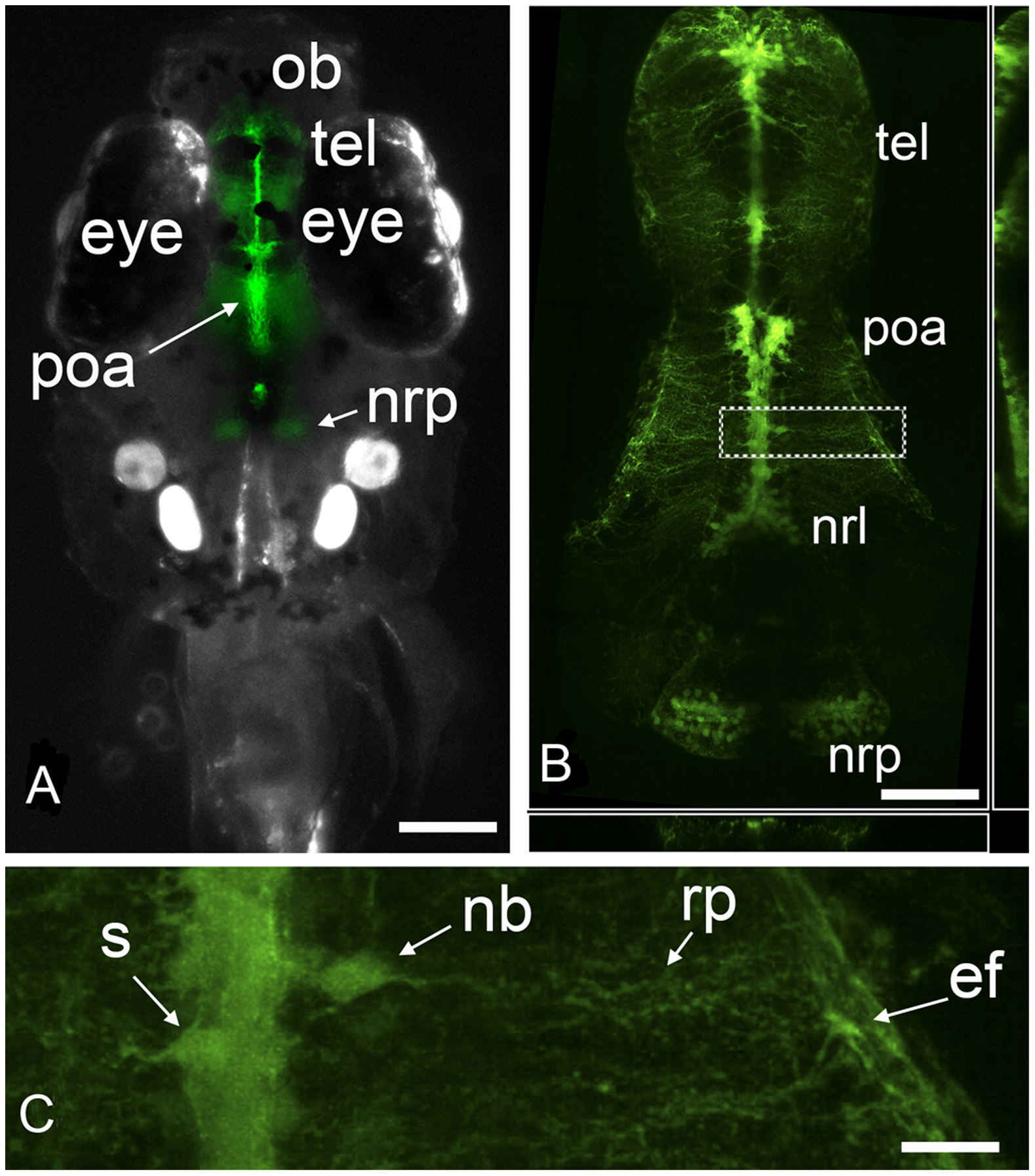
Fig. 1
Upon exposure of embryos to estradiol, the tg(cyp19a1b-GFP) zebrafish expresses GFP only in radial glial cells.
(a) Dorsal view of a zebrafish larva treated with 10 nM E2 showing that GFP signal is visible in the brain, notably in the telencephalon (tel), preoptic area (poa), and in the nucleus recessus posterioris (nrp) of the caudal hypothalamus; ob: olfactory bulb. (b) High resolution confocal image showing the RGCs in the telencephalon (tel), preoptic area (poa), nucleus recessus lateralis (nrl) and nucleus recessus posterioris (nrp) of the caudal hypothalamus. (c) High power view of the area shown in (b). Soma (s) are located along the midline except in the case of newborn cells (nb) undergoing migration (see Figure 2). RGCs have long cytoplasmic radial processes (rp) terminating by end-feet (ef) at the brain surface. (a) Bar = 200 μm; (b) Bar = 100 μm (c) Bar = 20 μm.