
|
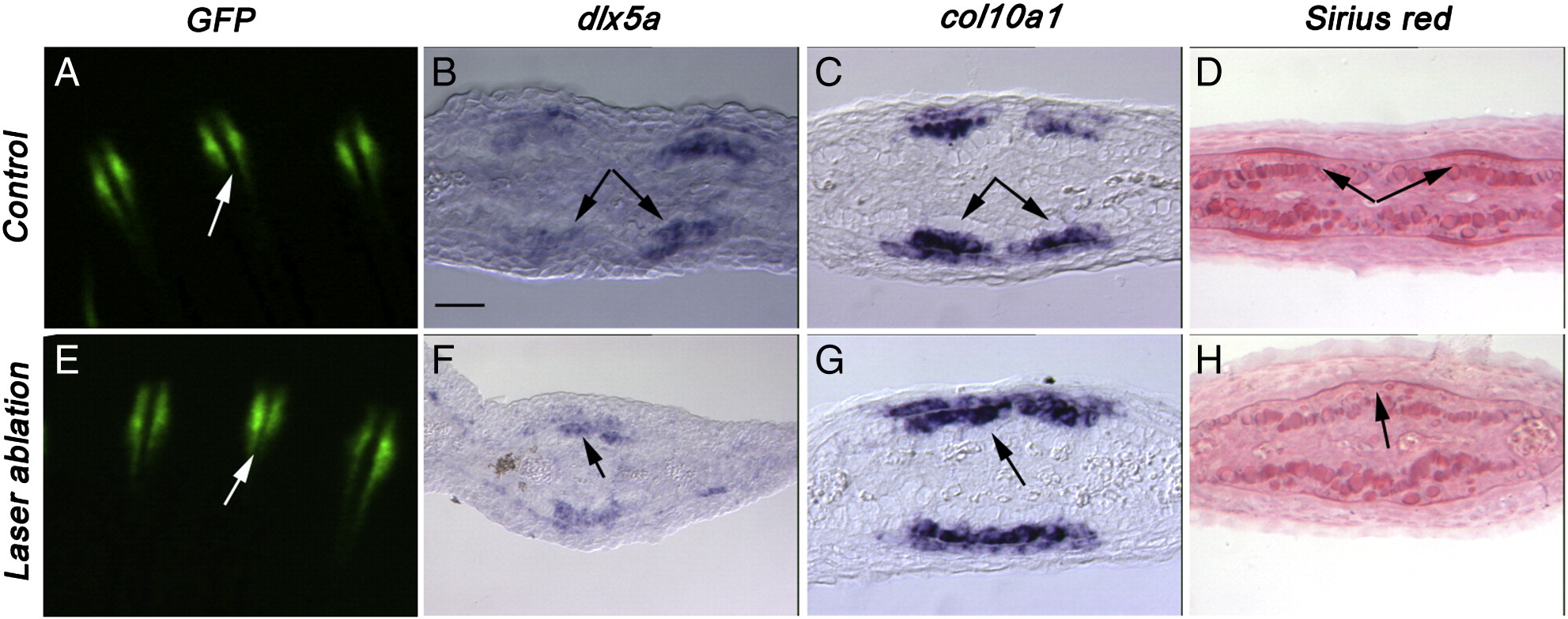
Fig. 6
Laser ablation induces a delay in the splitting of the domain of expression of dlx5a and col10a1. At 6 dpa 3 dpl, control (A, B, C and D) and laser-ablated fin rays (E, F, G and H) were observed for GFP expression (A and E) or collected for gene expression analysis (B, C, D and F) or picrosirius red staining (G and H) on transverse sections. In control fin regenerates, at 6 dpa, (A) GFP expression (B) dlx5a, (C) col10a1 and (D) picrosirius red staining are observed in two separate domains. In experimental fin rays, at 6 dpa, 3 days after ablation of the GFP positive cells, (E) GFP expression is starting to separate in two domains while (F) dlx5a, (G) col10a1 and (H) picrosirius red staining are still observed in a unique domain in each fin ray. Arrows indicate one or two domains of expression/staining per fin ray. Scale bar in panels B–H = 20 μm.
Reprinted from Developmental Biology, 365(2), Zhang, J., Jeradi, S., Strähle, U., and Akimenko, M.A., Laser ablation of the sonic hedgehog-a-expressing cells during fin regeneration affects ray branching morphogenesis, 424-433, Copyright (2012) with permission from Elsevier. Full text @ Dev. Biol.