
IMAGE
Fig. S7
- ID
- ZDB-IMAGE-120425-19
- Publication
- Sultana et al., 2008 - Zebrafish early cardiac connexin, Cx36.7/Ecx, regulates myofibril orientation and heart morphogenesis by establishing Nkx2.5 expression
- All Figures
- Figures for Sultana et al., 2008
Image
|
Figure Caption
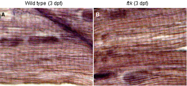
Fig. S7
Formation of striated myofibers in the skeletal muscles. (A and B) Hematoxylin/eosin staining of sagittal sections of trunk skeletal muscles. Wild-type (A) and ftk mutant (B) are shown at 3 dpf. A typical striated muscle structure was observed in both samples, indicating that ftk mutation did not affect the myofibril organization in the skeletal muscle.
Figure Data
Acknowledgments
This image is the copyrighted work of the attributed author or publisher, and
ZFIN has permission only to display this image to its users.
Additional permissions should be obtained from the applicable author or publisher of the image.
Full text @ Proc. Natl. Acad. Sci. USA