
|
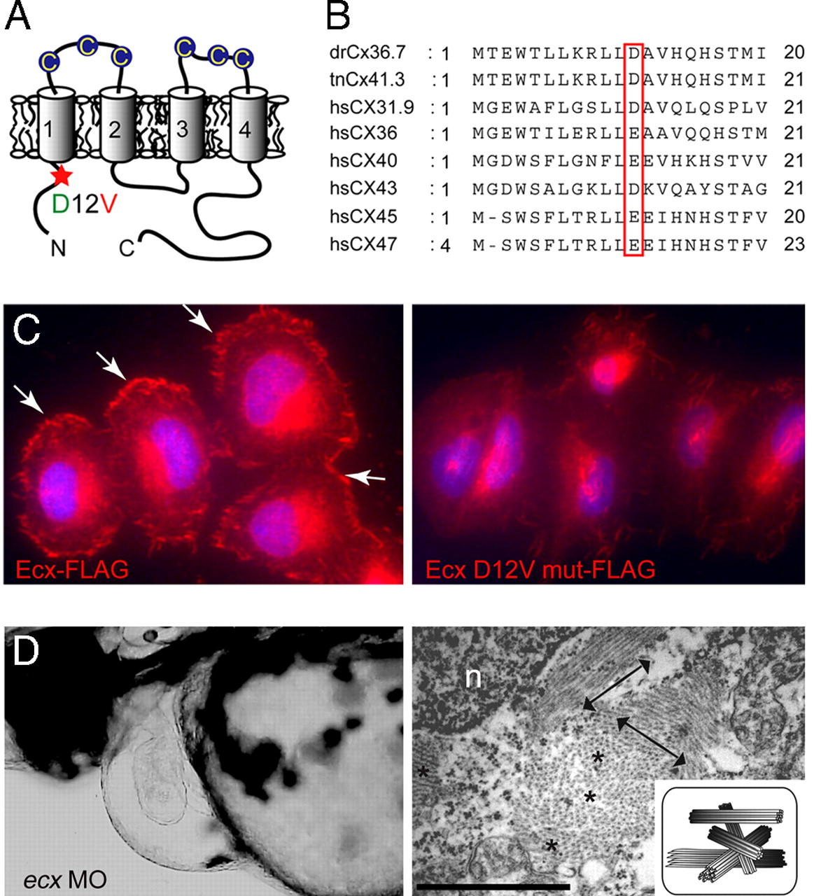
Fig. 4
ftk is a loss-of-function mutation of ecx/cx36.7. (A) Putative topology of Ecx protein. Extracellular cysteine residues are indicated by circles. The D12V mutation located in the N-terminal intracellular region of Ecx is indicated with a red asterisk. (B) Alignment of the N-terminal domain of zebrafish Ecx with Tetraodon Cx41.3 and six human connexins. (C) Subcellular localization of FLAG-tagged Ecx proteins in HeLa cells. Wild-type Ecx is localized at the cell surface (Left, arrows), whereas the mutant protein is not recruited to the membrane surface (Right). Nuclei were stained with Hoechst 33342 (blue). (D) Production of ftk phenotype with the ecx MO. (Left) Severe pericardiac edema and dilated heart chambers are observed in larvae injected with ecx MO. Lateral view at 56 hpf is shown. (Right) Disorganized myofibrils as seen in ftk mutants were also caused by the ecx MO. Double-headed arrows and asterisks indicate the myofibril bundles sectioned parallel and perpendicular to the long axis of the cells, respectively. (Scale bar: 500 nm.) n, nucleus. (Inset) Schematic of myofibril organization.