Fig. 5
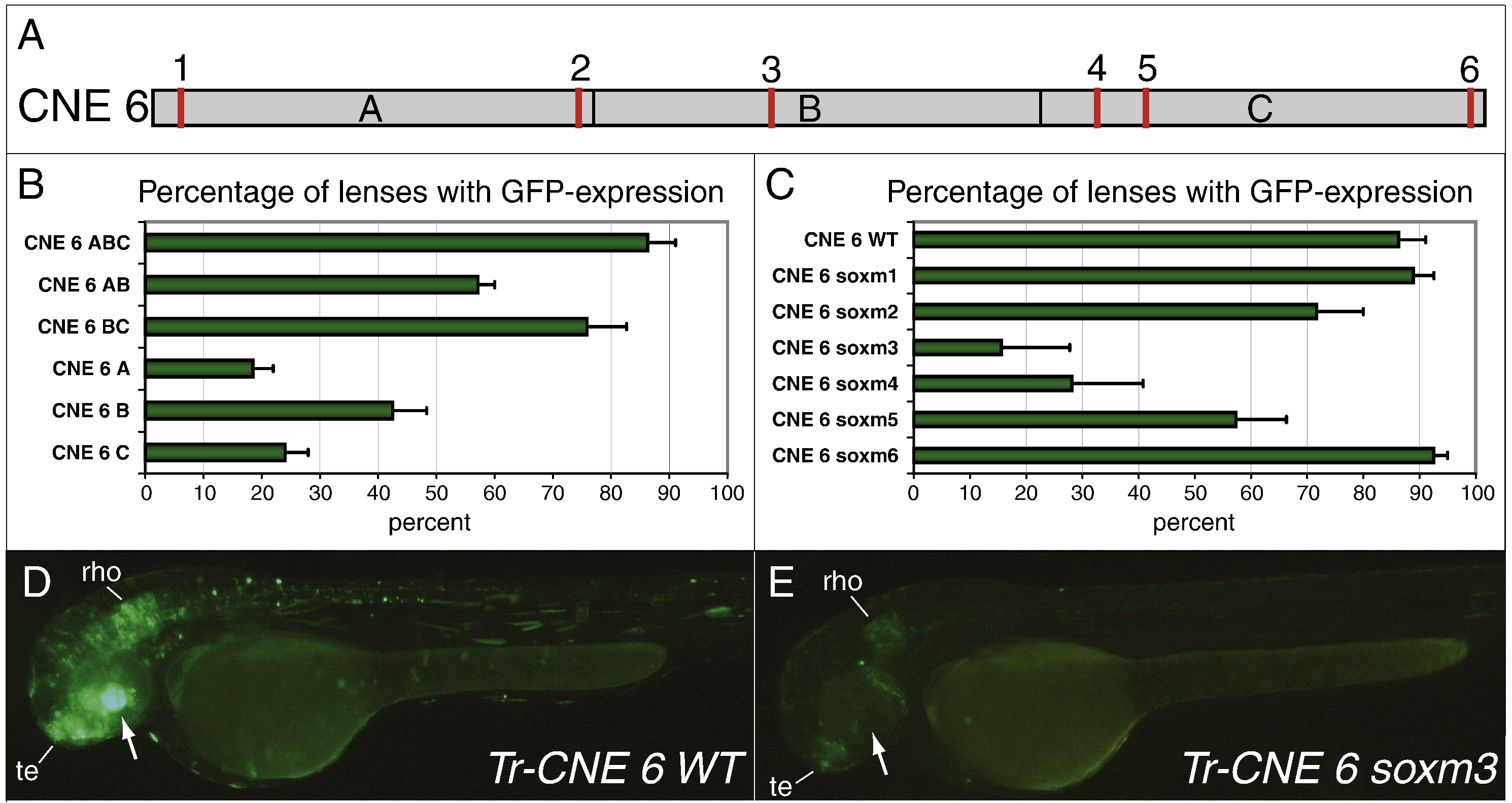
Dissection of the Fugu CNE 6 lens enhancer. (A) Schematic drawing of CNE 6 to indicate the subdivision into three parts (ABC) for the deletion analysis and the position of the six putative Sox binding sites (red) numbered 1–6. (B) Percentage of GFP-positive lenses in zebrafish injected with various deletion clones of Fugu CNE 6 (ABC = full-length CNE 6). (C) Percentage of GFP-positive lenses in zebrafish injected with different mutated versions of CNE 6. All 6 putative Sox sites were mutated separately (soxm1-6). (D,E) Transient GFP expression in 52 hpf-old zebrafish injected with the Fugu CNE 6 WT sequence (D) or a variant carrying a mutation in putative Sox site 3 (E). The lens is indicated by an arrow. Note that absence of lens expression in (E) correlates with lower expression levels in telencephalon (te) and rhombencephalon (rho). Tr (Takifugu rubripes, Fugu).
Reprinted from Developmental Biology, 365(1), Pauls, S., Smith, S.F., and Elgar, G., Lens development depends on a pair of highly conserved Sox21 regulatory elements, 310-318, Copyright (2012) with permission from Elsevier. Full text @ Dev. Biol.

