
Fig. S4
- ID
- ZDB-IMAGE-120405-93
- Publication
- Wang et al., 2012 - LAR Receptor Tyrosine Phosphatases and HSPGs Guide Peripheral Sensory Axons to the Skin
- All Figures
- Figures for Wang et al., 2012
|
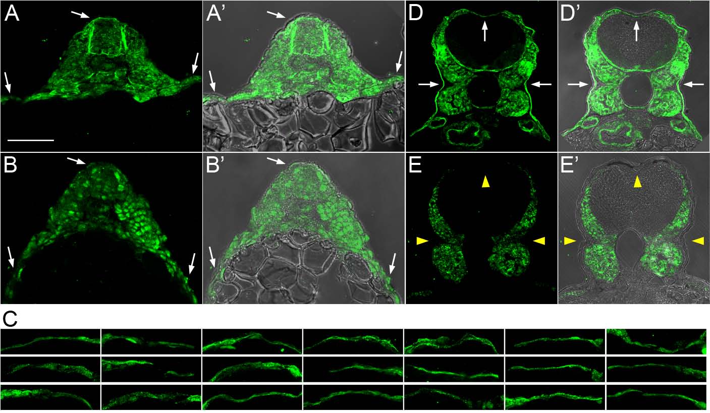
Fig. S4
Heparan Sulfate Staining in dackel Mutants and Siblings, Related to Figure 5
Cross-sections of anti-heparan sulfate (HS)-stained 18 to 20 somite-stage (SS) embryos (A-C) and 3 dpf larvae (D-E). At 18-20 SS, there was no morphological difference between dackel mutants and wildtype embryos. 23 embryos were stained and imaged, 5-6 of which were expected to be dackel mutants. Widespread HS staining was seen in all 23 embryos. Only one embryo appeared to have less extracellular HS staining (B) compared to others (e.g., A), but HS staining was nonetheless clearly seen around the yolk, which is surrounded by skin. This staining was not seen in controls lacking primary antibodies (not shown). (C) Representative regions of skin around the yolk from the other 21 embryos. (D) At 3 dpf, non-mutant siblings of dackel mutants displayed prominent staining in basement membranes, but HS staining was not detectable in basement membranes in dackel mutants (E). Arrows indicate skin, arrowheads indicate the absence of HS from skin in 3 dpf dackel mutants. n=6 for each 3 dpf group. Scale bar indicates 50μm.