
IMAGE
Fig. S3
- ID
- ZDB-IMAGE-120405-65
- Publication
- Miyake et al., 2012 - Neucrin, a novel secreted antagonist of canonical Wnt signaling, plays roles in developing neural tissues in zebrafish
- All Figures
- Figures for Miyake et al., 2012
Image
|
Figure Caption
Fig. S3
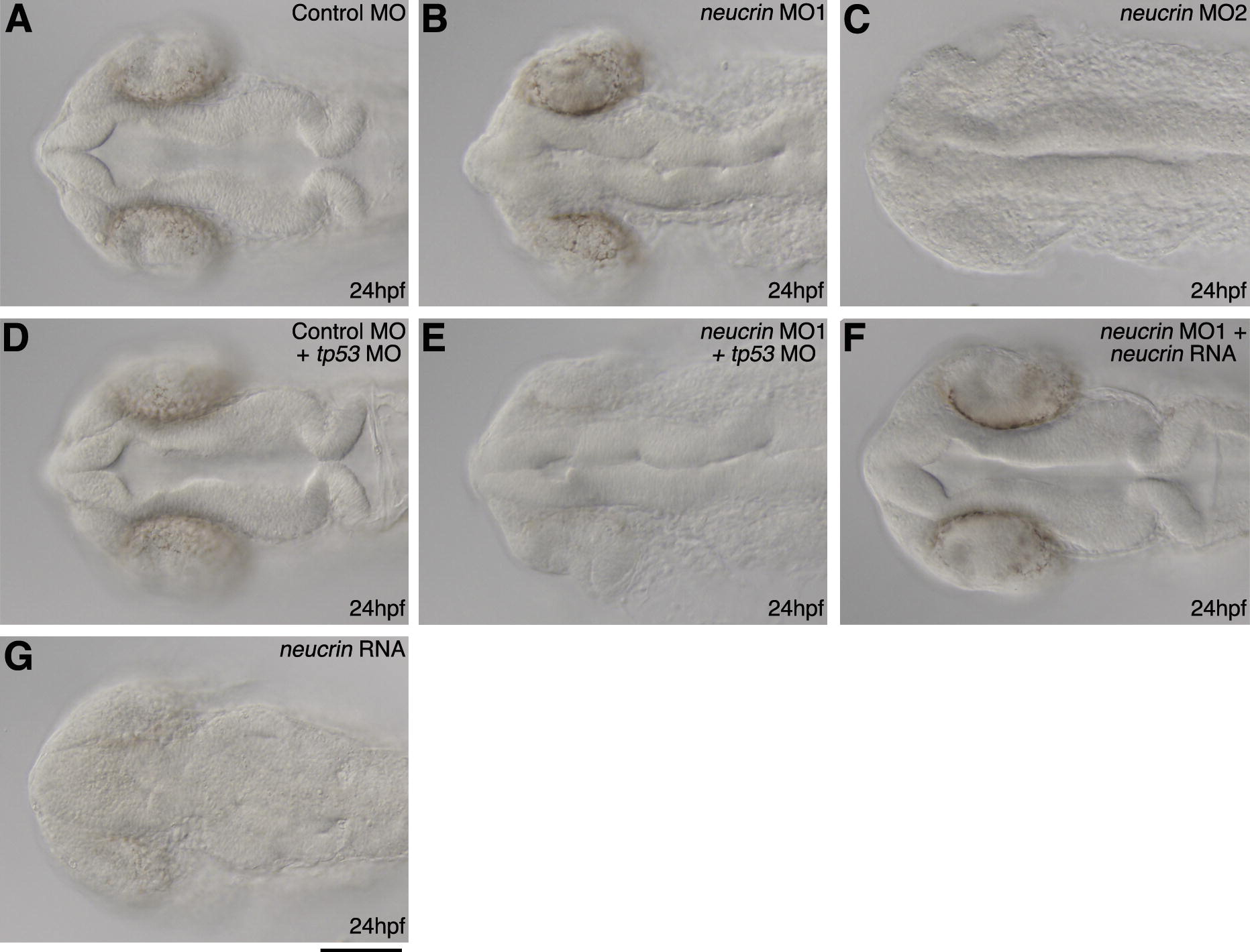
Morphology in the embryos injected with neucrin MOs at the 16-cell stage. Dorsal view of control MO-injected (A), neucrin MO1-injected (B), neucrin MO2-injected (C), tp53 MO- and control MO-injected (D), tp53 MO- and neucrin MO1-injected (E), neucrin MO1- and neucrin RNA-injected (F), or neucrin RNA-injected (G) embryos at 24 hpf are shown. Scale bar: 100 μm.
Figure Data
Acknowledgments
This image is the copyrighted work of the attributed author or publisher, and
ZFIN has permission only to display this image to its users.
Additional permissions should be obtained from the applicable author or publisher of the image.
Reprinted from Mechanisms of Development, 128(11-12), Miyake, A., Nihno, S., Murakoshi, Y., Satsuka, A., Nakayama, Y., and Itoh, N., Neucrin, a novel secreted antagonist of canonical Wnt signaling, plays roles in developing neural tissues in zebrafish, 577-590, Copyright (2012) with permission from Elsevier. Full text @ Mech. Dev.