
Fig. 7
- ID
- ZDB-IMAGE-120405-62
- Publication
- Miyake et al., 2012 - Neucrin, a novel secreted antagonist of canonical Wnt signaling, plays roles in developing neural tissues in zebrafish
- All Figures
- Figures for Miyake et al., 2012
|
Fig. 7
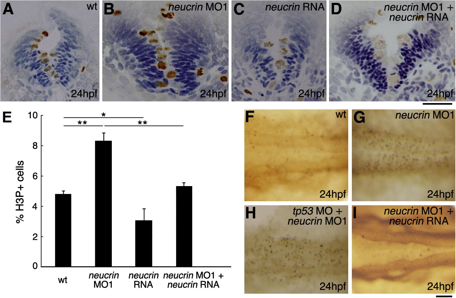
Comparison of cell proliferation and death in neucrin MO1-injected and neucrin RNA-injected embryos. (A-E) Wild-type embryos (A) and embryos injected with neucrin MO1 (B), neucrin RNA (C), or neucrin MO1 and neucrin RNA (D) were stained using an anti-phosphorylated Histone H3 antibody. (A-D) Panels show representative transverse sections of the hindbrain at 24 hpf. Brown and blue cells indicate the H3P-positive cells and H3P-negative cells, respectively. (E) The percentage of proliferating cells labeled with anti-H3P antibody in the hindbrain of wild-type embryos and embryos injected with neucrin MO1 or neucrin RNA or co-injected with neucrin MO1 and neucrin RNA. Results are the means ± S.D. for three embryos. The statistical significance of differences in mean values was assessed with the Student’s t-test. Asterisks indicate statistical significance compared with the wild type (*P < 0.05; **P < 0.01). (F-I) At 24 hpf, apoptotic cells in the hindbrain of wild-type embryos (F) and embryos injected with neucrin MO1 (G), neucrin MO1 and p53 MO (H), or neucrin MO1 and neucrin RNA (I) were marked via TUNEL labeling. Dorsal view with anterior to the left. Scale bar: 50 μm.
Reprinted from Mechanisms of Development, 128(11-12), Miyake, A., Nihno, S., Murakoshi, Y., Satsuka, A., Nakayama, Y., and Itoh, N., Neucrin, a novel secreted antagonist of canonical Wnt signaling, plays roles in developing neural tissues in zebrafish, 577-590, Copyright (2012) with permission from Elsevier. Full text @ Mech. Dev.