
IMAGE
Fig. 2
- ID
- ZDB-IMAGE-120405-57
- Genes
- Publication
- Miyake et al., 2012 - Neucrin, a novel secreted antagonist of canonical Wnt signaling, plays roles in developing neural tissues in zebrafish
- All Figures
- Figures for Miyake et al., 2012
Image
|
Figure Caption
Fig. 2
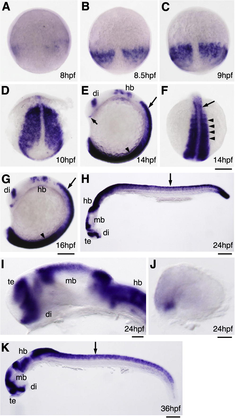
Expression of neucrin in zebrafish embryos. Expression pattern of neucrin in zebrafish embryos at the indicated stages detected by whole-mount in situ hybridization. (A-D, F) Dorsal view with anterior to the top. (E, G-K) Lateral view with anterior to the left and dorsal to the top. Long arrows and arrowheads indicate the spinal cord and somite, respectively. A short arrow in E indicates tightly clustered cell groups in the ventrorostral forebrain. di, diencephalon; hb, hindbrain; mb, midbrain; te, telencephalon. Scale bar: 100 μm in A-H, K; 25 μm in I, J.
Figure Data
Acknowledgments
This image is the copyrighted work of the attributed author or publisher, and
ZFIN has permission only to display this image to its users.
Additional permissions should be obtained from the applicable author or publisher of the image.
Reprinted from Mechanisms of Development, 128(11-12), Miyake, A., Nihno, S., Murakoshi, Y., Satsuka, A., Nakayama, Y., and Itoh, N., Neucrin, a novel secreted antagonist of canonical Wnt signaling, plays roles in developing neural tissues in zebrafish, 577-590, Copyright (2012) with permission from Elsevier. Full text @ Mech. Dev.