
|
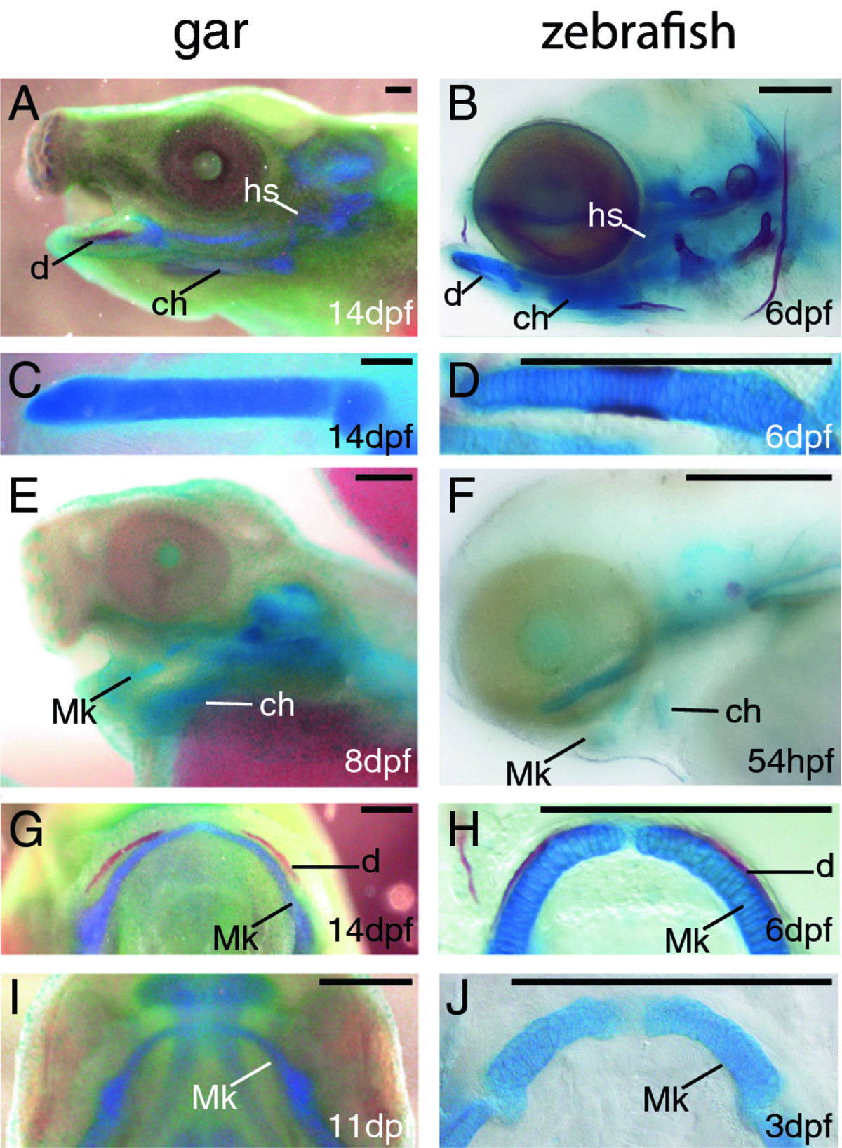
Fig. 2
Temporal series of skeletal preparations reveal early and late stages of cartilage and bone development in gar and zebrafish. A-J, Alcian blue (cartilage)/Alizarin red (mineralized bone) stained fish larvae. Lateral images of larval head skeletons show that many cartilage and a few bone elements were well-formed by 14 dpf in gar (A) and by 6 dpf in zebrafish (B). Dissected and flat-mounted ceratohyals of 14 dpf gar (C) and 6 dpf zebrafish (D) showed strong Alcian blue staining and distinct boundaries of the skeletal element, which are two features of well-developed cartilage. At earlier stages, more faint and diffuse Alcian blue staining was apparent in developing ceratohyal condensations of 8 dpf gar (E) and 54 hpf zebrafish (F). Ventral views show obvious Alizarin red staining of the dentary along anterolateral aspects of Meckel′s cartilage in the lower jaws of 14 dpf gar (G) and 6 dpf zebrafish (H). At earlier timepoints, no Alizarin red staining was visible in regions of the dentary in 11 dpf gar (I) and 3 dpf zebrafish (J). Scale bars: A-J = 0.25 mm. Abbreviations: ch = ceratohyal; d = dentary; dpf = days post-fertilization; hpf = hours post-fertilization; hs = hyosymplectic; Mk = Meckel′s.