
|
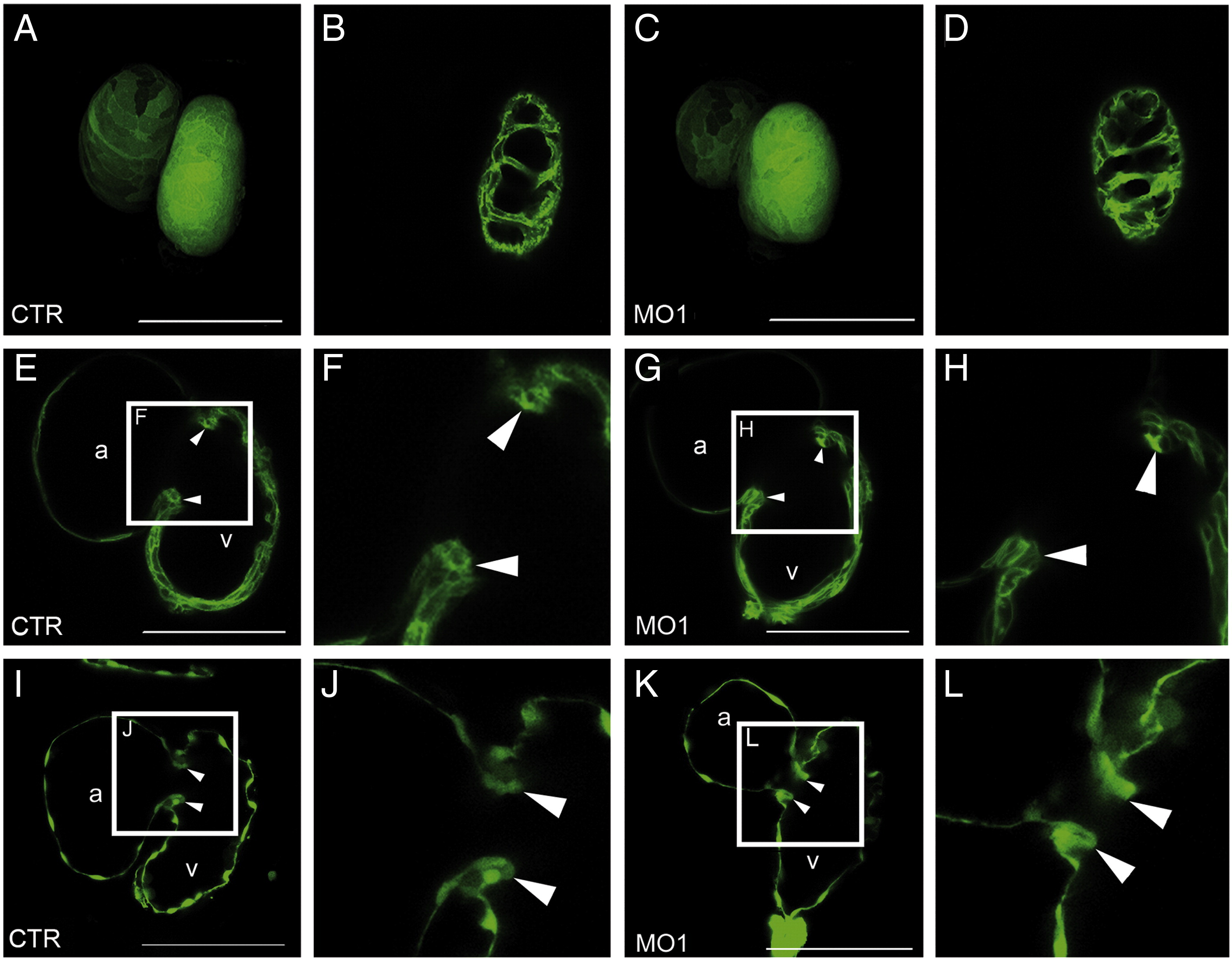
Fig. S8 Cardiac chamber dimensions and morphology of MO1-popdc2 morphants. Confocal analysis of 5 dpf (A–H) Tg(cmlc2:eGFP-ras)s883, and (I–L) Tg(flk1:eGFP)s843 hearts, which were injected with 1 ng (A,B,E,F,I,J) control (CTR), or (C,D,G,H,K,L) MO1-popdc2 morpholinos (MO1), respectively. (A,C) 3D visualization of atrial and ventricular chamber dimensions using confocal stacks. (B,D) Single images through the ventricular wall revealing normal trabeculation in popdc2 morphants displaying arrhythmia. The frames in panels (E,G,I,K) demarcate the area of magnification depicted in (F,H,J,L), respectively. No differences in morphology of the (E–H) AV myocardium or the (I–L) developing valve were observed between control and MO1-popdc2 morphants with rhythm defects. Abbreviations: a—atrium, v—ventricle. Scale bar = 100 μm.
Reprinted from Developmental Biology, 363(2), Kirchmaier, B.C., Poon, K.L., Schwerte, T., Huisken, J., Winkler, C., Jungblut, B., Stainier, D.Y., and Brand, T., The Popeye domain containing 2 (popdc2) gene in zebrafish is required for heart and skeletal muscle development, 438-450, Copyright (2012) with permission from Elsevier. Full text @ Dev. Biol.