
Image
|
Figure Caption
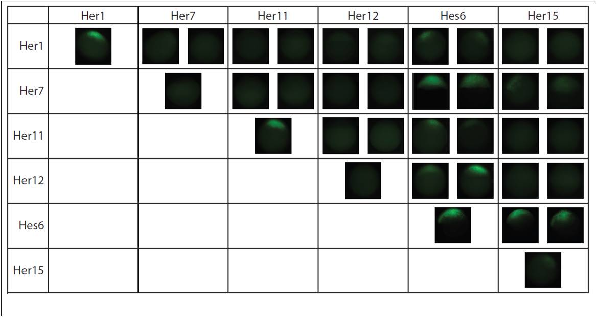
Fig. S3 Her dimers detected via BiFC. The chart shows the 10 of 21 Her combinations that dimerize in vivo. The first image in each cell is of the fusion to the N-terminal half of Venus YFP and the second is of the C-terminal half of Venus YFP for the Her protein indicated in the column header. Homodimers are observed along the diagonal. Some dimers appear weaker then others (Her1, Her1+Hes6, Her11+Hes6, Her7+Her15) and were detected only at higher expression levels.
Acknowledgments
This image is the copyrighted work of the attributed author or publisher, and
ZFIN has permission only to display this image to its users.
Additional permissions should be obtained from the applicable author or publisher of the image.
Full text @ Development