
|
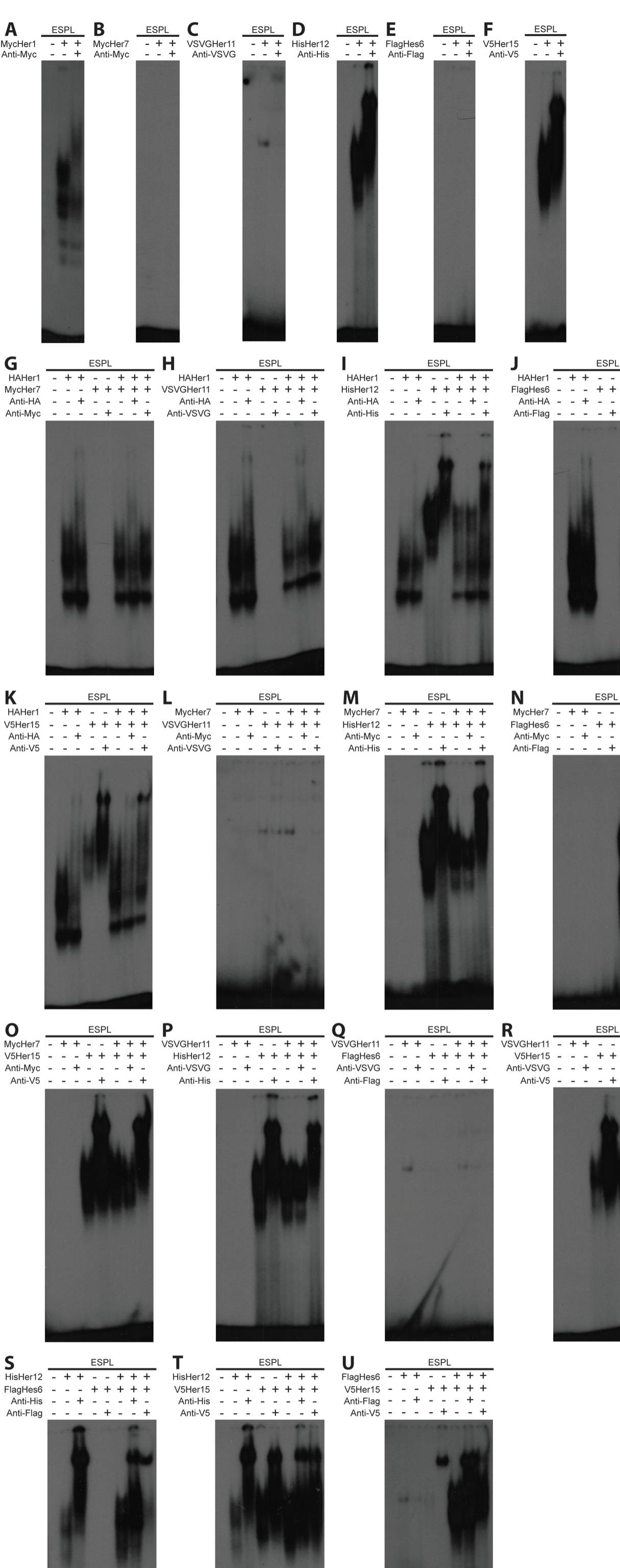
Fig. S2 Her dimers detected via EMSA. (A-F) Homodimer analysis for Her1 (A), Her7 (B), Her11 (C), Her12 (D), Hes6 (E) and Her15 (F). Only three our of six Her proteins form detectable homodimers. It is possible that Her11 forms a homodimer, but DNA binding is much weaker than the others and was not present in every gel. (G-U) All combinations of heterodimers: for Her1+Her7 (G), Her1+Her11 (H), Her1+Her12 (I), Her1+Hes6 (J), Her1+Her15 (K), Her7+Her11 (L), Her7+Her12 (M), Her7+Hes6 (N), Her7+Her15 (O), Her11+Her12 (P), Her11+Hes6 (Q), Her11+Her15 (R), Her12+Hes6 (S), Her12+Her15 (T), Hes6+Her15 (U). For Her1+Her12 (I), Her1+Her15 (K) and Her12+Her15 (T), the formation of heterodimers was being assayed for two proteins that form homodimers. In each of these cases, the pattern of dimerization appeared to be additive between the two homodimers, and no novel band or gel shift was detected.