
Fig. 3
- ID
- ZDB-IMAGE-120316-3
- Genes
- Antibodies
- Publication
- Zannino et al., 2012 - Olig2-expressing hindbrain cells are required for migrating facial motor neurons
- All Figures
- Figures for Zannino et al., 2012
|
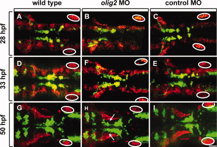
Fig. 3 olig2 MO does not disrupt lateral line migration. All panels show dorsal views of whole embryos, anterior to the left. Elavl labeling is shown in red and Tg(isl1:GFP)rw0 reporter expression is green. A–C: 28-hpf embryos. D–F: 33-hpf embryos. G–I: 50-hpf embryos. A–F show projections of z-stack images in both red and green channels. The red channel in G–I is a single z-plane, focused on the lateral line, whereas the green channel is a z-stack image projection of the green channel. Circles mark the posterior lateral line primordia. Arrows indicate lagging facial motor neurons in olig2 MO-injected embryos. Scale bar = 30 μm.