
Fig. 1
- ID
- ZDB-IMAGE-120315-20
- Publication
- Clendenon et al., 2012 - Zebrafish cadherin-11 participates in retinal differentiation and retinotectal axon projection during visual system development
- All Figures
- Figures for Clendenon et al., 2012
|
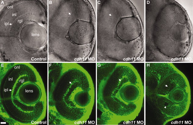
Fig. 1
Eyes of cdh11 morphants are small and lamina organization is disrupted at 2 days postfertilization (dpf). A–H: Zebrafish embryos were injected with either standard control morpholino oligonucleotide (MO; control, A and E) or cdh11 MO (B–D,F–H). A–D: Ventral views eyes of living embryos were imaged using differential interference contrast microscopy (A–D). E–H: BODIPY-ceramide labeled living 2 dpf embryos were imaged using confocal microscopy. Anterior is up and lateral is right. All the major layers characteristic of the adult retina are seen in control embryos, including the retinal ganglion cell layer (rgl), inner plexiform layer (ipl; arrow), the inner nuclear layer (inl), and the outer nuclear layer (onl). B–D,F–H: Increasing severity of Cdh11 loss-of-function phenotype were observed: B and F, in slightly affected embryos, eyes were similar sizes but retinal lamina were reduced; C and G, in moderately affected embryos, the eyes were smaller in size, the inner plexiform layer was sparse, the distance between the inner plexiform layer and the lens was not uniform, and the outer nuclear layer was sparse or absent; D and H, eyes and lenses of severely affected embryos were small, rosettes were evident (arrowhead) and laminae nearly absent. Scale bar = 20 μm.