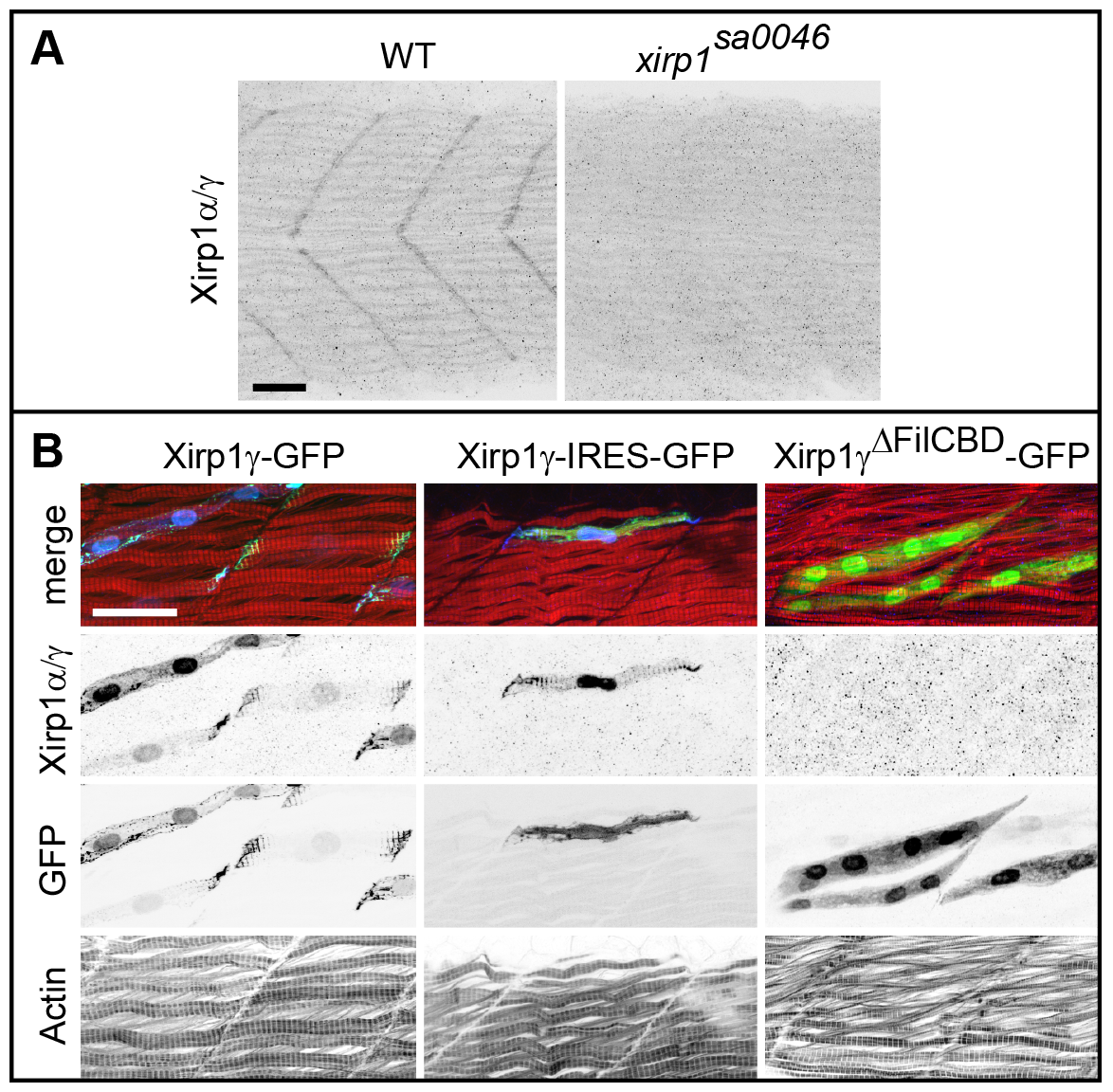
Fig. 8
Characterization of the P47 antibody.
(A) Myotendinous junction labeling by the P47 antibody which recognizes Xirp1α/γ is absent in xirp1sa0046 mutant embryos at 24 hpf. This staining was performed upon mild fixation. (B) Clonally expressed Xirp1γ is sensitively detected upon standard fixation conditions by the P47 antibody within somitic muscle tissue. Truncated Xirp1γΔFilCBD-GFP which lacks the Filamin C binding domain including the P47 epitope is not detected by the antibody. Under standard fixation conditions, Xirp1α/γ is not detected at the myotendinous junctions. Green: GFP or Xirp1γ-GFP fusion protein; red: Actin; blue: Xirp1α/γ. All scale bars: 25 μm.

