
Fig. 3
- ID
- ZDB-IMAGE-120228-20
- Publication
- Nicoli et al., 2012 - miR-221 Is Required for Endothelial Tip Cell Behaviors during Vascular Development
- All Figures
- Figures for Nicoli et al., 2012
|
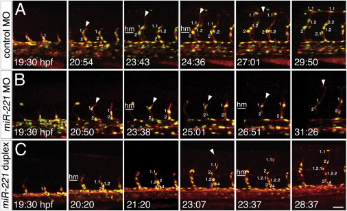
Fig. 3
miR-221 Regulates Migration and Proliferation of Tip Cells (A–C) Still images from time-lapse analysis of (A and B) Tg(fli1a:negfp)y7;(kdrl:ras-cherry)s916 or Tg(fli1a:negfp)y7;(kdrl:tagrfap-caax)is19 embryos. Time (hpf) is noted in the bottom left hand corner. Nuclei are numbered; successive numbers indicate new cells that migrated from the dorsal aorta, decimals indicate daughter cells arising from cell division. Lateral views, dorsal is up, anterior to the left. Horizontal white line denotes horizontal myoseptum (hm). (A) Embryo injected with 10 ng control MO. (B) Embryo injected with 10 ng miR-221 MO. (C) Embryo injected with 500 µM miR-221 duplex. Scale bar is 25 μm. See mmc3VIDEO, mmc4VIDEO and mmc5VIDEO.
Reprinted from Developmental Cell, 22(2), Nicoli, S., Knyphausen, C.P., Zhu, L.J., Lakshmanan, A., and Lawson, N.D., miR-221 Is Required for Endothelial Tip Cell Behaviors during Vascular Development, 418-429, Copyright (2012) with permission from Elsevier. Full text @ Dev. Cell