
|
Fig. 1
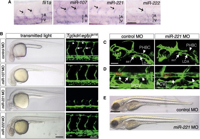
miR-221 Is Required for Vascular Development (A) Whole-mount in situ hybridization of embryos at 48 hr postfertilization (hpf). Dorsal aorta and posterior cardinal vein are indicated by a bracket and A or V, respectively. Arrows indicate intersegmental blood vessels (ISV). Scale bar = 50 μm. (B) Left column, transmitted light images of embryos at 30 hpf injected with indicated MO. Scale bar = 250 μm. Right column, confocal micrographs of trunk vessels at 30 hpf in Tg(kdrl:egfp)la116 embryos injected with indicated MO. ISVs indicated by arrows and DLAV by arrowheads. Scale bar = 50 μm. (C) Tg(kdrl:egfp)la116 embryos at 27 hpf injected with 10 ng control or miR-221 MO. aa1, aortic arch 1; PHBC, primordial hindbrain channel; LDA, lateral dorsal aorta. Scale bar = 50 μm. (D) Tg(fli1a:egfp)y1 embryos at 5 days postfertilization (dpf) injected as in (C). DA, dorsal aorta; PCV, posterior cardinal vein; position of thoracic duct is indicated by arrows. Scale bar = 25 μm. (E) Transmitted light images of larvae at 5 dpf injected as in (C). Scale bar = 500 μm. All panels are lateral views, dorsal is up, anterior to the left.
Reprinted from Developmental Cell, 22(2), Nicoli, S., Knyphausen, C.P., Zhu, L.J., Lakshmanan, A., and Lawson, N.D., miR-221 Is Required for Endothelial Tip Cell Behaviors during Vascular Development, 418-429, Copyright (2012) with permission from Elsevier. Full text @ Dev. Cell