
|
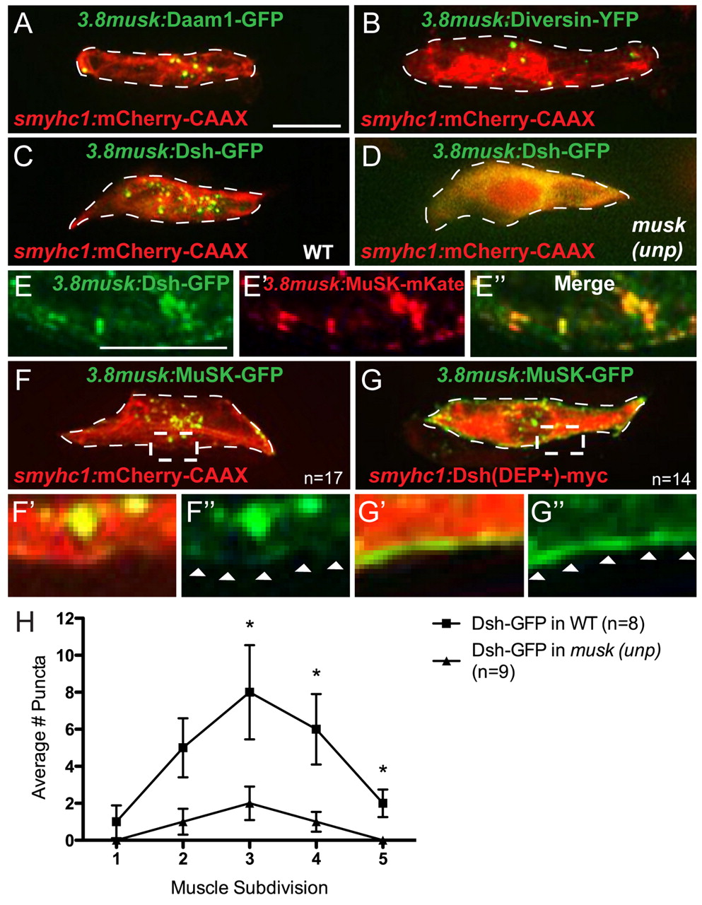
Fig. 5
Localization of noncanonical PCP proteins to the center of muscle cells requires MuSK, and vice versa. (A,B) Daam1-GFP (green) (A) and Diversin-YFP (green) (B) under the 3.8musk promoter localize to centrally enriched puncta in muscle cells co-expressing mCherry-CAAX under the smyhc1 promoter (red). (C) Dsh-GFP under the 3.8musk promoter (green) localizes to centrally enriched puncta in fibers co-expressing mCherry-CAAX (red). (D) The punctate localization of Dsh-GFP (green) is lost in musk (unp) mutant muscle cells (mCherry-CAAX in red). (E-E3) Magnified view of center of muscle cell showing colocalization of Dsh-GFP (green) and MuSK-mKate (red), both driven by the 3.8musk promoter. (F-F3) MuSK-GFP under the 3.8musk promoter (green) localizes to centrally enriched puncta in muscle cells co-expressing mCherry-CAAX (red). F2 and F3 show magnified views of the region marked with dashed box in F. (G-G3) MuSK-GFP localizes to the membrane of muscle cells co-expressing Dsh(DEP+)-Myc. G2 and G3 show magnified views of the region marked with dashed box in G. (H) Quantification of reduction in centrally localized Dsh-GFP puncta in musk (unp) mutants (P values for muscle subdivisions 1-5: 0.49, 0.09, 0.05, 0.04, 0.02, respectively). *P<0.05. Error bars represent s.e.m. Dashed lines encircle a single muscle cell. Arrowheads in F3 and G3 indicate cell membrane. Scale bars: 10 μm.