
Fig. S3
- ID
- ZDB-IMAGE-120216-73
- Genes
- Antibodies
- Publication
- Yoruk et al., 2012 - Ccm3 functions in a manner distinct from Ccm1 and Ccm2 in a zebrafish model of CCM vascular disease
- All Figures
- Figures for Yoruk et al., 2012
|
Fig. S3
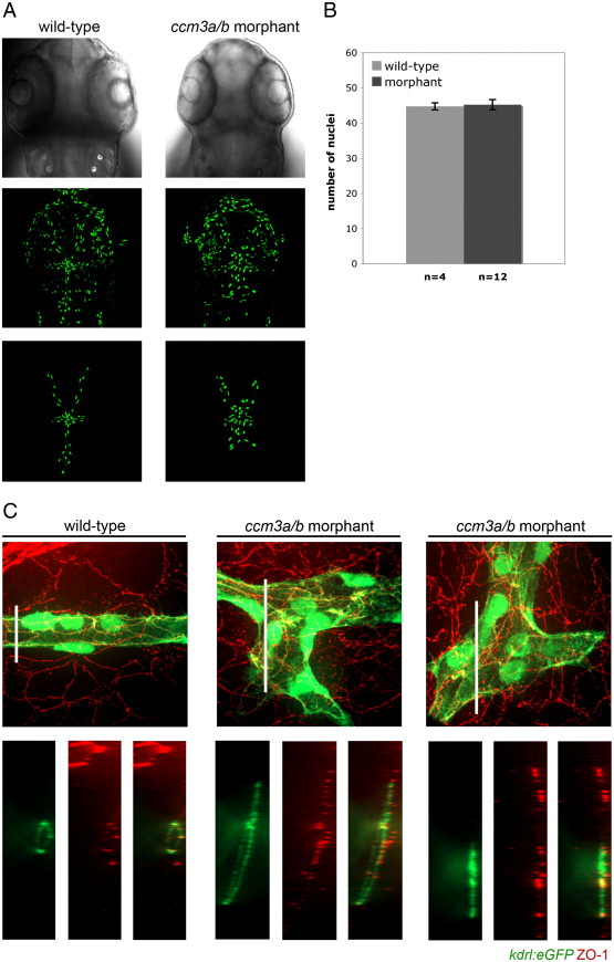
Cranial vasculature enlargements seen in ccm3a/b morphants is not due to overproliferation of endothelial cells in the affected vessels. (A) Confocal z-stack projections of dorsal head vasculature in wildtype and ccm3a/b morphant embryos in the Tg(fli1:nlsEGFP) background at 54 hpf. (B) Endothelial cell nuclei were quantified in ccm3a/b morpholino injected (0.75 ng) and non-injected mesencephalic veins using the Tg(fli1:nlsEGFP) line. There was no statistically significant difference between the endothelial cell numbers in morphants compared to wildtype (p-value = 0.78). (C) Tg(kdrl:EGFP) embryos at 54 hpf were stained with mouse anti-ZO-1, and confocal images were analyzed for ZO-1 expression (red), and blood vessels (green). Cross-sections of the z-stack projections, as indicated by the white bars, revealed that wildtype mesencephalic veins (MsVs) were properly lumenized with strong ZO-1 expression at tight junctions along the circumference of the vessels. ccm3a/b morphant MsVs were enlarged, and in many instances failed to lumenize. ZO-1 expression in the morphants appeared disorganized and showed decreased co-localization with the vasculature (green).
Reprinted from Developmental Biology, 362(2), Yoruk, B., Gillers, B.S., Chi, N.C., and Scott, I.C., Ccm3 functions in a manner distinct from Ccm1 and Ccm2 in a zebrafish model of CCM vascular disease, 121-131, Copyright (2012) with permission from Elsevier. Full text @ Dev. Biol.