Fig. S1
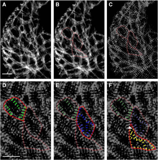
Illustration of method for measuring cardiomyocyte cell size and myofibril content. (A) To measure cell surface area and total Z-band size, we begin by visualizing cardiomyocyte boundaries, marked here by phalloidin staining in a ventral view of the ventricle, arterial pole at the top, at 48 hpf. (B) Cortical actin filaments are substantially thicker than the actin filaments in myofibrils that cross the cell. We therefore trace cell contours (red outlines) based on thick cortical phalloidin staining and then measure the area of the resulting polygons, as in our prior work (Auman et al., 2007). (C) Red polygons are overlaid on the corresponding image of anti-α-actinin staining in the same ventricle to provide visualization of myofibril Z-bands within individual cells. (D) We trace line segments (green lines) corresponding to individual Z-bands that appear in a series of at least three parallel bands. Because the ventricular myocardial wall is only one cell thick at the stages examined (Peshkovsky et al., 2011) (Fig. S6), we can assign observed Z-bands reliably to individual cells. (D) We trace line segments (green lines) corresponding to individual Z-bands that appear in a series of at least three parallel bands. Because the ventricular myocardial wall is only one cell thick at the stages examined (Peshkovsky et al., 2011) (Fig. S6), we can assign observed Z-bands reliably to individual cells. (E, F) The process of tracing line segments (blue and yellow lines) continues in each individual cell. When Z-bands cross borders of neighboring cells, the line segments are subdivided and the contribution of each portion is assigned to its respective cell (white arrowhead). Scale bars are 10 μm.
Reprinted from Developmental Biology, 362(2), Lin, Y.F., Swinburne, I., and Yelon, D., Multiple influences of blood flow on cardiomyocyte hypertrophy in the embryonic zebrafish heart, 242-253, Copyright (2012) with permission from Elsevier. Full text @ Dev. Biol.

