
Fig. 4
- ID
- ZDB-IMAGE-120203-7
- Publication
- Li et al., 2012 - Augmenter of Liver Regeneration (alr) Promotes Liver Outgrowth during Zebrafish Hepatogenesis
- All Figures
- Figures for Li et al., 2012
|
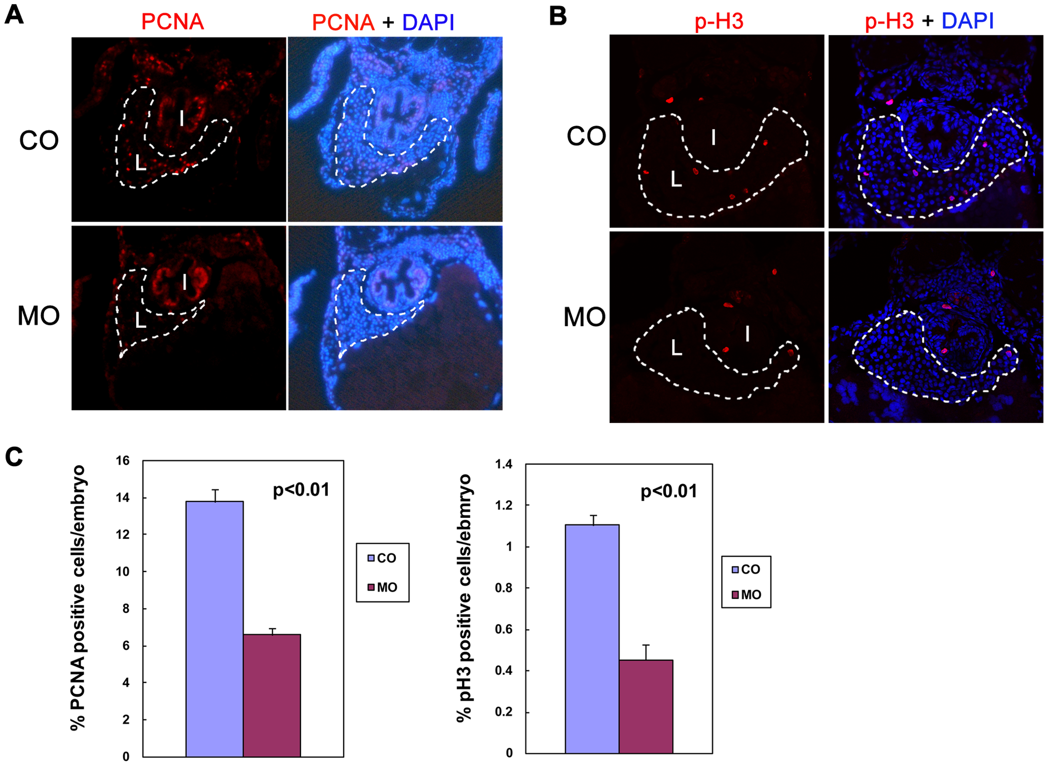
Fig. 4
Knockdown of alr reduces hepatocyte proliferation.
A & B. Hepatocyte proliferation demonstrated by immunofluorescent staining of proliferation markers in 4 dpf embryos: proliferating cell nuclear antigen (PCNA) (A) and phosphor-histone 3 (p-H3) (B). The sections were counterstained with DAPI to label nucleus. PCNA and p-H3 staining is co-localized with DAPI, indicative of nucleus staining. Both PCNA and p-H3 staining showed a significantly reduced hepatocyte proliferation in morphants without affecting proliferation in other tissues such as intestine. I: intestine; L: liver. Dash line circles the boundary of liver. C. Quantification of hepatocyte proliferation. Percentage of PCNA positive hepatocytes in liver is reduced from 13.8% in CO to 6.6% in MO. Percentage of p-H3 positive hepatocytes in liver is reduced from 1.1% in CO to 0.45% in MO. Values are means ± standard deviation (SD). Hepatocytes were counted based on cell morphology.