
|
Fig. S8
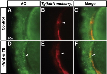
Increased Wnt signaling at the TB stage induces cell death at the same anatomical position of the anterior LPM as endothelial cells. (A–C) A representative HCSE. (D–F) A representative GFP + embryo after increasing Wnt signaling at the TB stage. To examine the position of apoptotic cells relative to endothelial cells of the LPM, hemizygous Tg(hsp70l:wnt8a-GFP)w34 fish were crossed to hemizygous Tg(kdrl:mCherry)ci5 fish as in Fig. 7. In a transgenic embryo after Wnt signaling was increased at the TB stage, there are apoptotic cells (arrows in D and F) adjacent to the endothelials in the anterior LPM (arrows in B and E). The HCSE does not have a significant amount of apoptotic cells in the same region of the anterior LPM. The anatomical position of these dying cells and endothelial cells is reminiscent of the more dorsal position of CMs relative to the endothelial cells reported in Bussmann et al. (2007). Arrow in B indicates endothelial cells that are more medial endocardial precursors. Images are lateral views with anterior to the top and dorsal to the left.
Reprinted from Developmental Biology, 361(2), Dohn, T.E., and Waxman, J.S., Distinct phases of Wnt/β-catenin signaling direct cardiomyocyte formation in zebrafish, 364-76, Copyright (2012) with permission from Elsevier. Full text @ Dev. Biol.