
|
Fig. 5
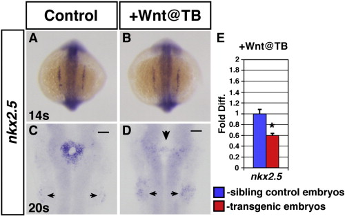
Increased Wnt signaling at the TB stage causes a loss of nkx2.5 expression after the 14 somite stage. (A,C) HCSEs. (B,D) GFP + sibling embryos. Expression of nkx2.5 is normal in embryos at the 14 s stage after increasing Wnt signaling at the TB stage (B; 100%, n = 27), compared to HCSEs (A; n = 38). (C) nkx2.5 expression in HCSEs (n = 21). (D) Expression of nkx2.5 is lost by 20 s (100% had severely reduced nkx2.5 expression, n = 27) in GFP + embryos after heat-shock at the TB stage. There are diffuse nkx2.5 expressing cells at the midline (large arrowhead). Posterior pharyngeal arch staining appears increased in embryos with increased Wnt signaling at the TB stage (small arrowheads). (E) qPCR analysis of nkx2.5 expression indicates that total nkx2.5 expression is reduced in 20–22 s stage GFP + embryos after increasing Wnt signaling at the TB stage.
Reprinted from Developmental Biology, 361(2), Dohn, T.E., and Waxman, J.S., Distinct phases of Wnt/β-catenin signaling direct cardiomyocyte formation in zebrafish, 364-76, Copyright (2012) with permission from Elsevier. Full text @ Dev. Biol.