
|
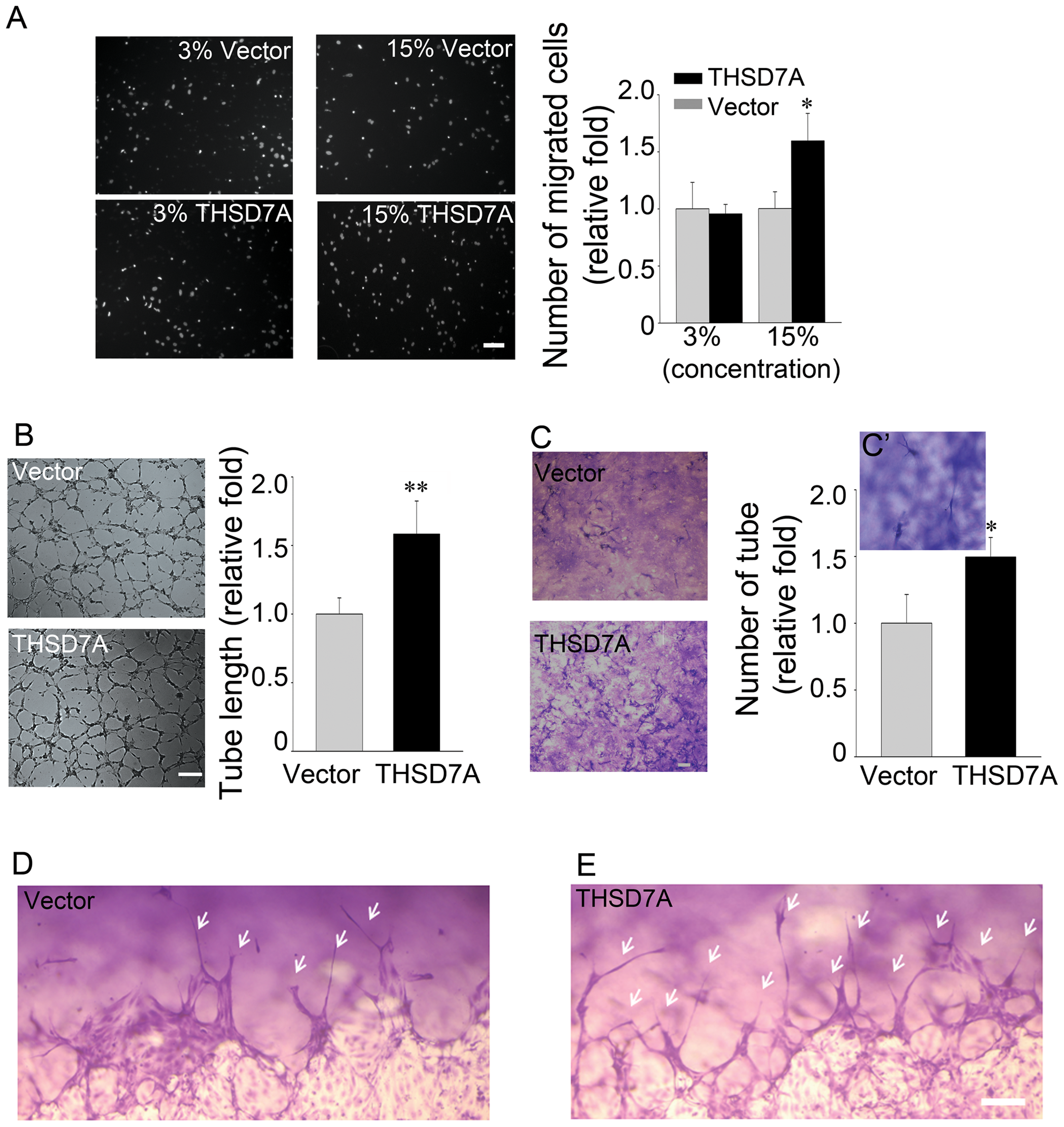
Fig. 4
Soluble THSD7A accelerates endothelial cell migration, tube formation and sprouting.
A. The motility of HUVEC was evaluated by transwell migration assay in M200 medium. HUVEC were loaded in the upper insert and soluble THSD7A or control medium were added to a 3% or 15% final concentration into the lower chamber. B. Two-dimensional tube formation was performed in the presence or absence of soluble THSD7A on Matrigel. C. Three-dimensional tube formation was performed in the presence or absence of soluble THSD7A on type I collagen as an assay for sprouting. C2. Enlarged image of tubes. Bar represents 10 μm. Each experiment was repeated at least three independent times. *P<0.05 vs. vector. D–E. Side views of the sprouting assay. Arrows indicate tubes. Bar represents 100 μm.