
IMAGE
Fig. S3
- ID
- ZDB-IMAGE-120131-54
- Publication
- Du et al., 2012 - Differential regulation of epiboly initiation and progression by zebrafish Eomesodermin A
- All Figures
- Figures for Du et al., 2012
Image
|
Figure Caption
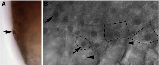
Fig. S3 Eomesa expression in the EVL. Antibody staining for Eomesa (A) Lateral view of embryo showing Eomesa in EVL nuclei, arrow. (B) Marginal view of embryo with Eomesa expression in EVL (arrows) but not YSL nuclei (arrowheads). Black dashed lines outline selected EVL cell boundaries.
Acknowledgments
This image is the copyrighted work of the attributed author or publisher, and
ZFIN has permission only to display this image to its users.
Additional permissions should be obtained from the applicable author or publisher of the image.
Reprinted from Developmental Biology, 362(1), Du, S., Draper, B.W., Mione, M., Moens, C.B., and Bruce, A.E., Differential regulation of epiboly initiation and progression by zebrafish Eomesodermin A, 11-23, Copyright (2012) with permission from Elsevier. Full text @ Dev. Biol.