
Fig. 2
- ID
- ZDB-IMAGE-120125-8
- Publication
- Powell et al., 2011 - Jamb and jamc are essential for vertebrate myocyte fusion
- All Figures
- Figures for Powell et al., 2011
|
Fig. 2
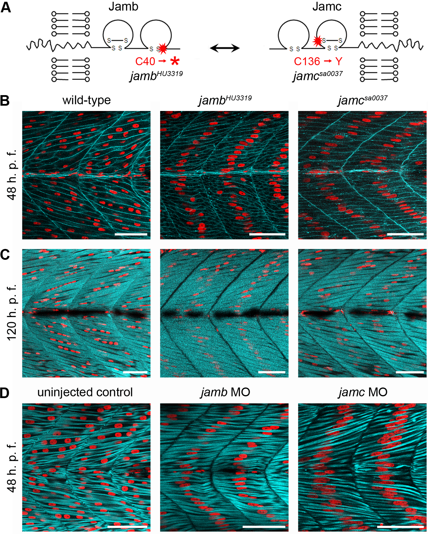
jamb and jamc are essential for myocyte fusion in vivo.
(A) Schematics of Jamb and Jamc extracellular proteins. Red stars denote sites of mutation in HU3319 and sa0037 alleles. (B–C) Confocal microscopy images of fast-twitch muscle in wild-type, jambHU3319, and jamcsa0037 48 h. p. f. (B) and 120 h. p. f. (C) Embryos labelled with membrane targeted RFP (mRFP, cyan; B) or phalloidin-Alexa488 (cyan; C) and DAPI (red) show overabundant, mononuclear myofibres in both mutants. (D) Confocal microscopy images of uninjected, jamb, and jamc translation-blocking morpholino-injected wild-type embryos, stained with DAPI (red) and phalloidin-Alexa488 (cyan) to stain F-actin in fast muscle fibres. The morpholino-injected embryos replicate the jam mutants′ phenotype. Myotomes 12–13 shown, anterior left. Scale bars represent 50 μm.