
IMAGE
Fig. S10
- ID
- ZDB-IMAGE-111220-20
- Publication
- Kimmel et al., 2011 - Requirement for Pdx1 in specification of latent endocrine progenitors in zebrafish
- All Figures
- Figures for Kimmel et al., 2011
Image
|
Figure Caption
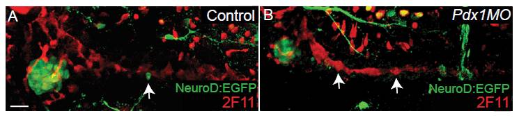
Fig. S10 Larval duct morphology. Confocal projection of 6 days post fertilization (dpf) TgBAC(NeuroD:EGFP)nl1 embryos immunostained for green fluorescent protein (GFP) and 2F11. In control embryos (A), 2F11 positive cells surround GFP+ islet cells and extend into the pancreatic tail. Single NeuroD+ cells can be found in the pancreas tail (arrow). (B) pdx1 morphant embryos, with fewer GFP+ cells in the islet, have similar 2F11 expression around the islet and in the pancreatic tail (arrows). Lateral view. Scale bar = 30 μM.
Acknowledgments
This image is the copyrighted work of the attributed author or publisher, and
ZFIN has permission only to display this image to its users.
Additional permissions should be obtained from the applicable author or publisher of the image.
Full text @ BMC Biol.