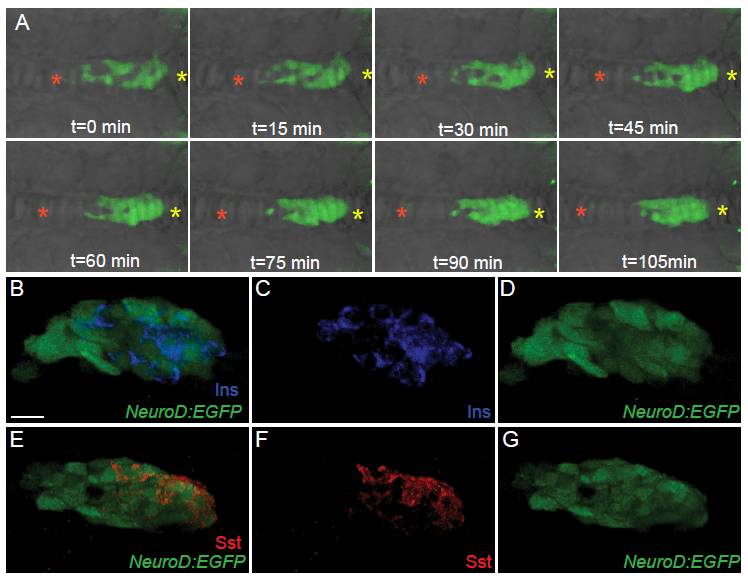
Fig. S3 NeuroD: enhanced green fluorescent protein (EGFP) cells are endocrine precursors. (A) TgBAC(NeuroD:EGFP)nl1 embryos, mounted dorsal side up, imaged by confocal timelapse microscopy starting at 19 h post fertilization (hpf). Images were captured every 15 min. Asterisks indicate fixed points of the embryo determined from the bright field image. Anterior is to the left. (B-G) Confocal image projection of 24 hpf TgBAC(NeuroD:EGFP)nl1 embryo immunostained for green fluorescent protein (GFP) and insulin (Ins) (B-D) and GFP and somatostatin (Sst) (E-G), showing overlap of NeuroD:EGFP expression with islet hormones in a subset of cells. All are ventral view. Scale bar = 15 μM.
Image
Figure Caption
Acknowledgments
This image is the copyrighted work of the attributed author or publisher, and
ZFIN has permission only to display this image to its users.
Additional permissions should be obtained from the applicable author or publisher of the image.
Full text @ BMC Biol.

