
|
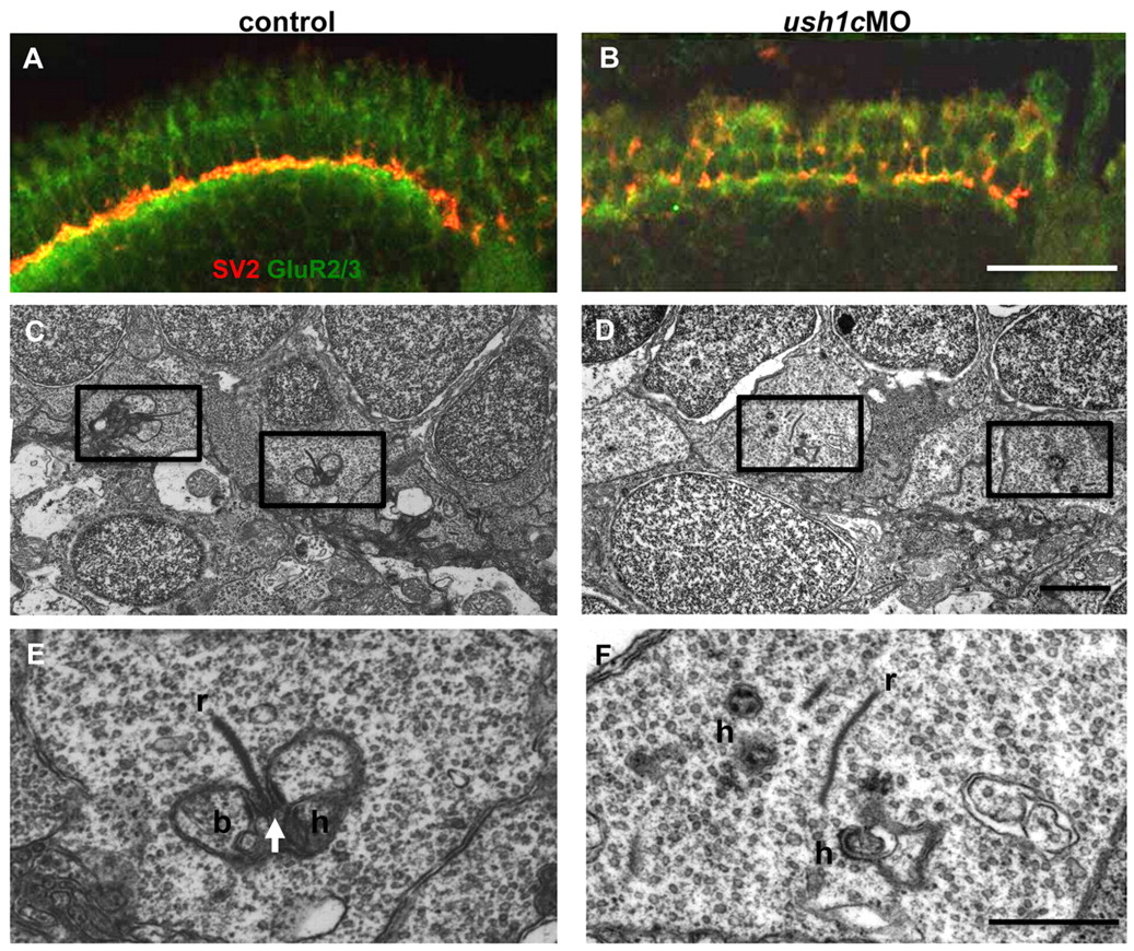
Fig. 7 Synaptic maturation is impaired in ush1c morphants. (A,B) Anti-SV2 (red) and -GluR2/3 (green) antibodies mark the pre-and postsynaptic regions of the OPL, respectively. Tightly associated localization of these factors is apparent in the control retina (A), and is notably disrupted in the morphant retina (B) at 5 dpf. (C–F) Electron micrographs of cone pedicles in 6 dpf control and morphant larvae. In control retinas (C,E), the presence of multiple triads (boxed areas), consisting of a synaptic ribbon (r), arciform density (arrow) and postsynaptic processes from bipolar (b) and horizontal (h) cells, indicate normal synaptic maturation. An enlargement of a triad is shown in E. In morphant retinas (D,F), fewer triads are present (boxed areas in D; enlarged in panel F), and floating ribbons (r) and distant postsynaptic processes (h) are observed in cone pedicles. Scale bars: 20 μm (A,B); 1 μm (C,D); 500 nm (E,F).