
IMAGE
Fig. S5
- ID
- ZDB-IMAGE-111215-16
- Publication
- Oehlers et al., 2011 - The inflammatory bowel disease (IBD) susceptibility genes NOD1 and NOD2 have conserved anti-bacterial roles in zebrafish
- All Figures
- Figures for Oehlers et al., 2011
Image
|
Figure Caption
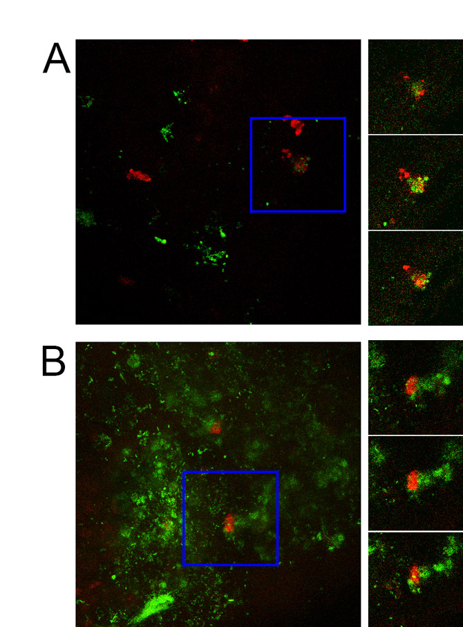
Fig. S5 Confocal imaging demonstrates phagocytosis of GFP-labelled S. enterica by zebrafish neutrophils. Images on left are maximum intensity Z-projections of 1 dpi (A) and 2 dpi (B) Tg(lyzC:dsRed)50 embryos injected with 200 CFU of GFP-tagged S. enterica into the yolk sac at 2 dpf. Blue box demarcates area of interest. Images on right show three consecutives slices from confocal stack from the area of interest.
Acknowledgments
This image is the copyrighted work of the attributed author or publisher, and
ZFIN has permission only to display this image to its users.
Additional permissions should be obtained from the applicable author or publisher of the image.
Full text @ Dis. Model. Mech.