
Fig. S3
- ID
- ZDB-IMAGE-111215-14
- Publication
- Oehlers et al., 2011 - The inflammatory bowel disease (IBD) susceptibility genes NOD1 and NOD2 have conserved anti-bacterial roles in zebrafish
- All Figures
- Figures for Oehlers et al., 2011
|
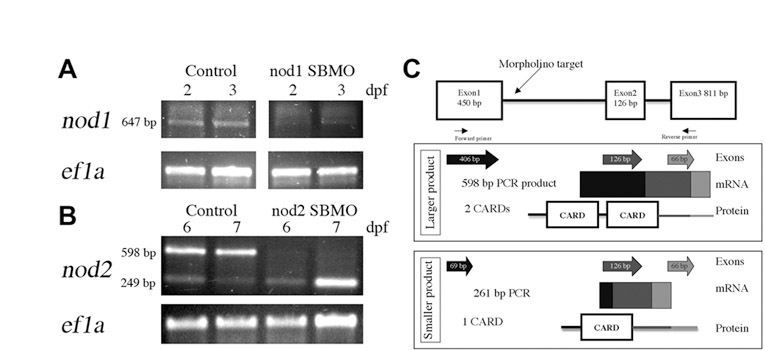
Fig. S3 Confirmation of Nod knockdown in zebrafish larvae. RT-PCR detection of (A) nod1 and (B) nod2 transcripts following MO-mediated depletion from 2 and 3 dpf (A) and 6 and 7 dpf (B) larvae. Ef1a was used as a control. (C) In silico analysis of predicted proteins derived from zebrafish Nod2 splice variants. The genomic structure with morpholino target and primer binding sites is shown at top. Exons amplified by RT-PCR are depicted as individual arrows and as a continuous amplicon of mRNA. Predicted protein structure is illustrated with shading indicating the contribution of each exon to the predicted domains.