
|
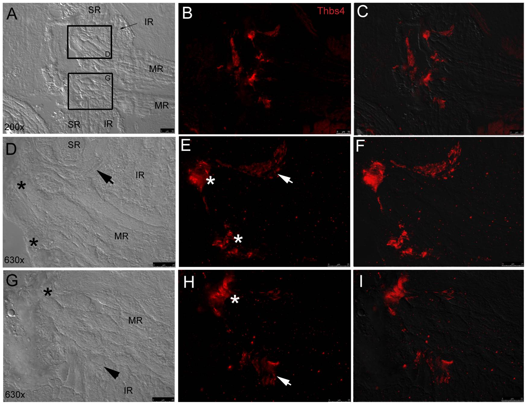
Fig. 9 Fluorescent immunohistochemistry shows thbs-4 enrichment in EOM origin tendons.
Low magnification (20x objective) DIC image (A) displays pairs of MR, SR, and IR converging at their common origin points with fluorescent thbs-4 (red) overlay (B) and fluorescent/DIC merged image (C). High magnification (63x) DIC images (D,G) show SR, IR, and MR attaching to bone with MR tendons (*) and IR tendons (arrows) marked. IR tendons marked with thbs-4 (E,F,H,I) are observed sandwiched at the furthest proximal point of the IR bordered by the MR and SR on either side. Thbs-4 is expressed in MR tendons (*) at their most proximal point as they insert on bone at their origin.