
Image
|
Figure Caption
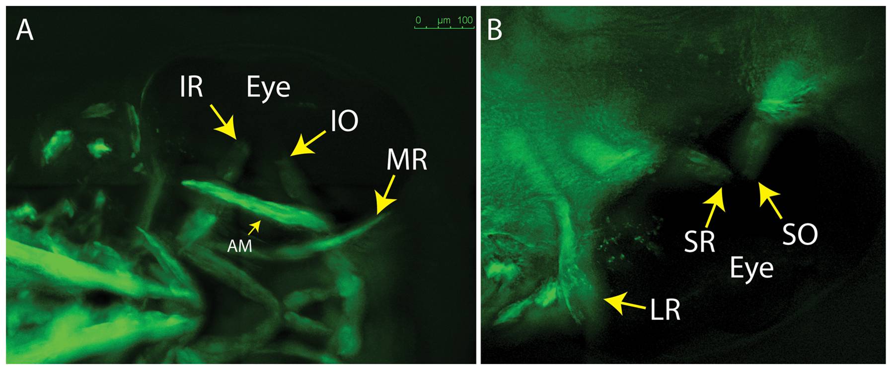
Fig. 3 5 dpf embryos showing lack of overlap for SR/SO and IR/IO at their insertion sites (arrows).
(A) ventral; (B) dorsal. Images reflect maximum projections following deconvolution of macroscopic Z-stacks. IR = inferior rectus; IO = inferior oblique; MR = medial rectus; AM = adductor mandibulae (jaw muscle – thin arrow); LR = lateral rectus; SR = superior rectus; SO = superior oblique.
Acknowledgments
This image is the copyrighted work of the attributed author or publisher, and
ZFIN has permission only to display this image to its users.
Additional permissions should be obtained from the applicable author or publisher of the image.
Full text @ PLoS One