
|
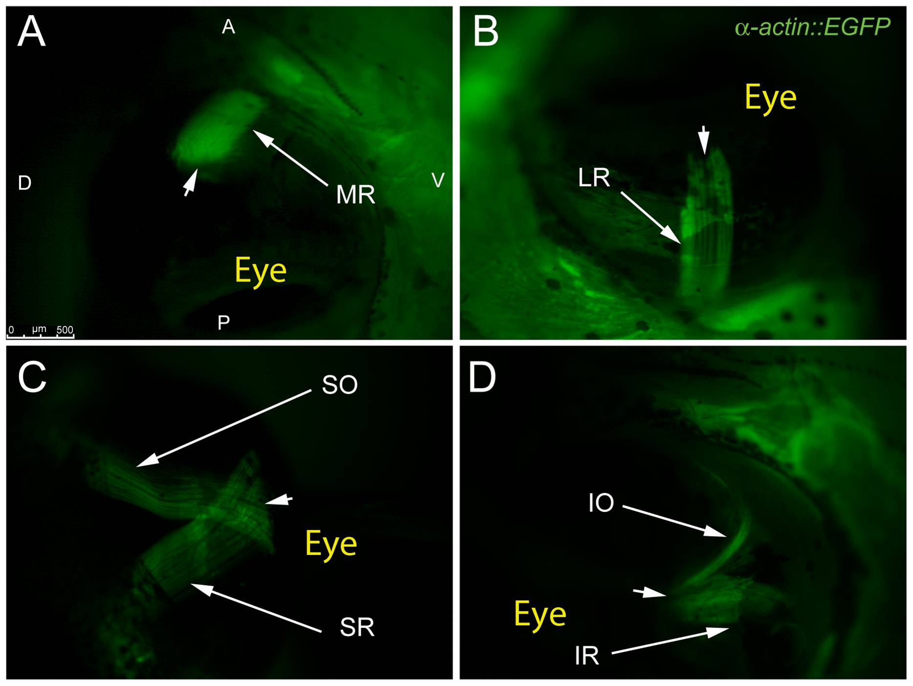
Fig. 2 Adult transgenic zebrafish expressing GFP under the control of the α-actin muscle protein promoter allow for clear visualization of all 6 EOMs using epifluorescent stereomicroscopy.
The MR and LR (A,B) are shown running parallel to the long axis of the fish and inserting on the anterior and posterior sides of the eye respectively. The SO and SR course from their respective rostral and caudal origin points to insert on the dorsal side of the eye with significant overlap of fibers (C). The IO and IR mirror the SO and SR as they insert onto the ventral side of the eye. Arrowheads mark scleromuscular insertion sites located at the scleral-corneal (SC) junction. Muscle origins are deep within the orbit and are not visible. The anterior (A), posterior (P), dorsal (D), and ventral (V) directions are noted on frame A and apply to frames B–D.