
|
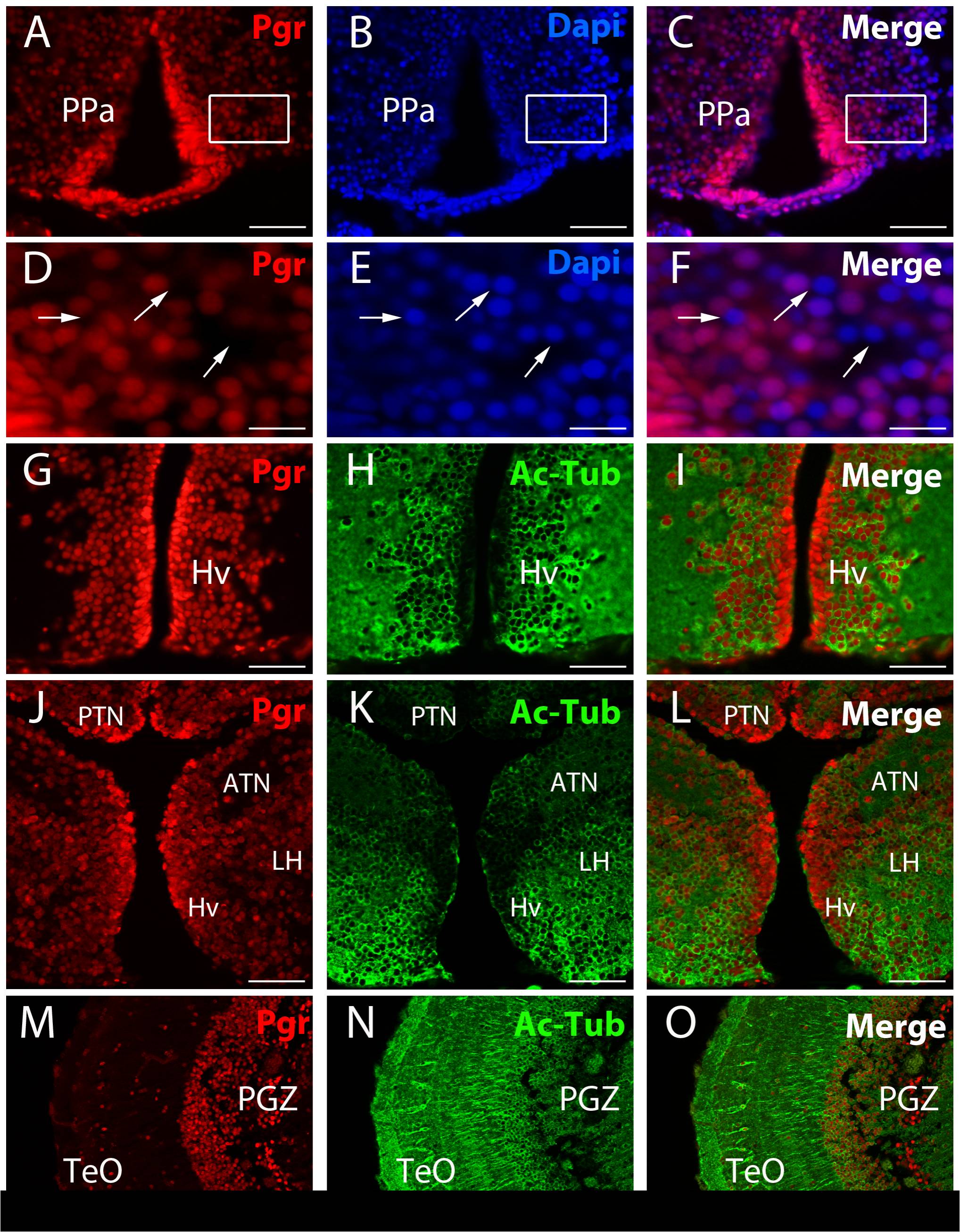
Fig. 2 Immunolocalisation of Pgr expressing neurons in adult zebrafish forebrain.
A to F: Pgr immunohistochemistry (red) with DAPI nuclei staining (blue) showing that not all nuclei express Pgr (arrows). D, E and F correspond to high magnifications of the area framed in A, B and C. Arrows point to nuclei that do not express Pgr. G to O: Pgr (red) and acetylated-tubulin (green) staining showing that numerous Pgr positive cells correspond to acetylated-tubulin positive neurons. Note that cells lining the ventricle are strongly positive for the Pgr antibodies, but are not stained by acetylated-tubulin. ATN: anterior tuberal nucleus; Hv: ventral zone of the periventricular hypothalamus; LH: lateral hypothalamic nucleus; PGZ: periventricular gray zone of the optic tectum; PPa: anterior part of the preoptic area; TeO: Optic tectum. Scale bars: 15 μm (D, E and F), 50 μm (A, B, C, G, H, I, J, K and L), 100 μm (M, N and O)