
Fig. 6
- ID
- ZDB-IMAGE-111213-14
- Publication
- Li et al., 2011 - Qilin is essential for cilia assembly and normal kidney development in zebrafish
- All Figures
- Figures for Li et al., 2011
|
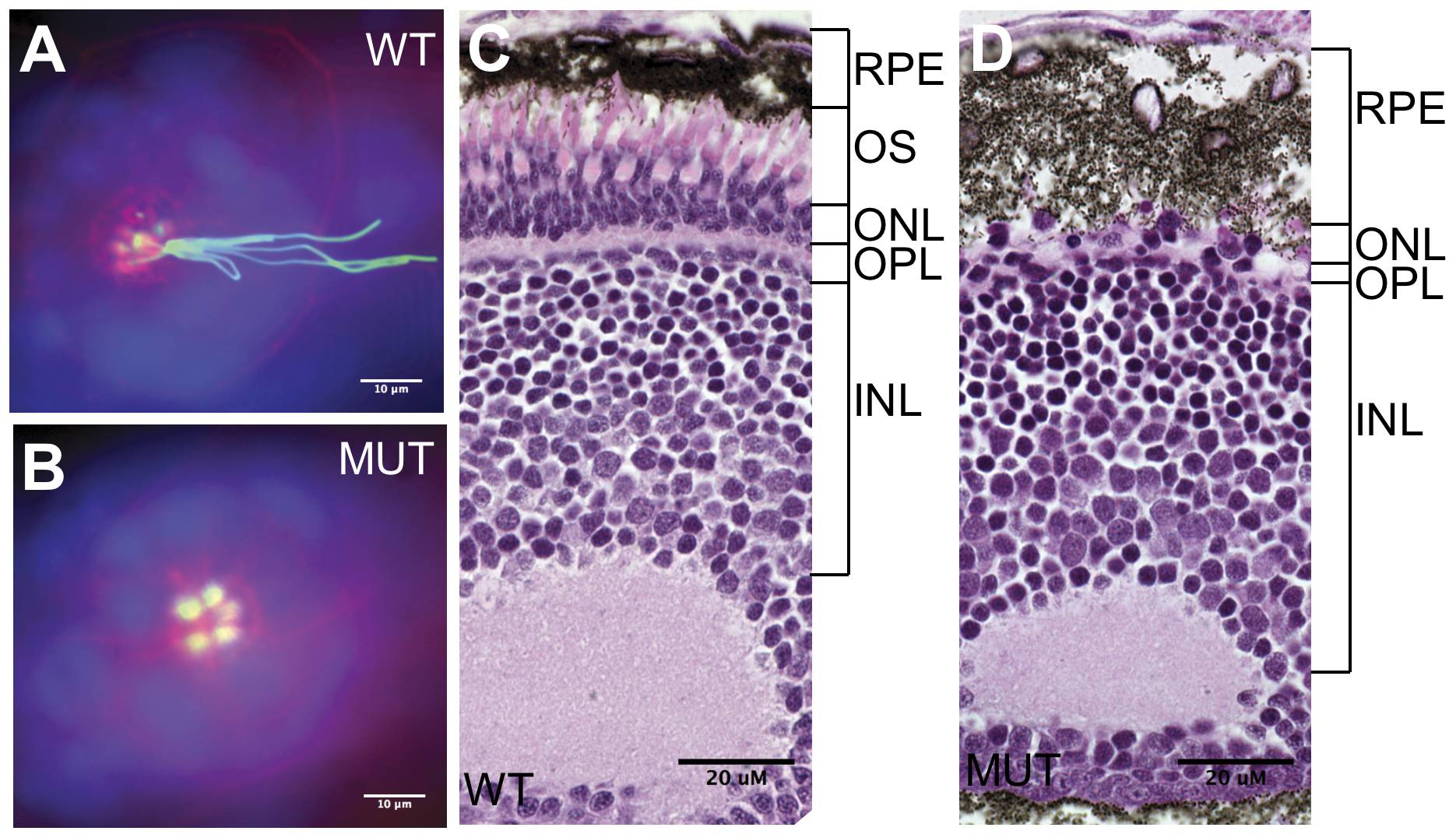
Fig. 6 Sensory cilia in qilin hi3959A mutants are defective.
(A–B) Epifluorescent projections showing the lateral line organ in a wild type (A) and a mutant embryo (B) at 3 dpf. Embryos were stained with rodamine-phalloidin (red), anti- acetylated tubulin (green) and DAPI (blue). Scale bars: 10 μm. (C–D) The absence of the outer segment of photoreceptors in the eye as shown through cross sections of a wild type (C) and a mutant embryo (D) at 5 dpf. WT: wild type; MUT: mutant; RPE: retinal pigment epithelium; OS: outer segment; ONL: the outer nuclear layer; INL: the inner nuclear layer; OPL: the outer plexiform layer. Scale bars: 20 μm.