
Fig. 5
- ID
- ZDB-IMAGE-111213-13
- Publication
- Li et al., 2011 - Qilin is essential for cilia assembly and normal kidney development in zebrafish
- All Figures
- Figures for Li et al., 2011
|
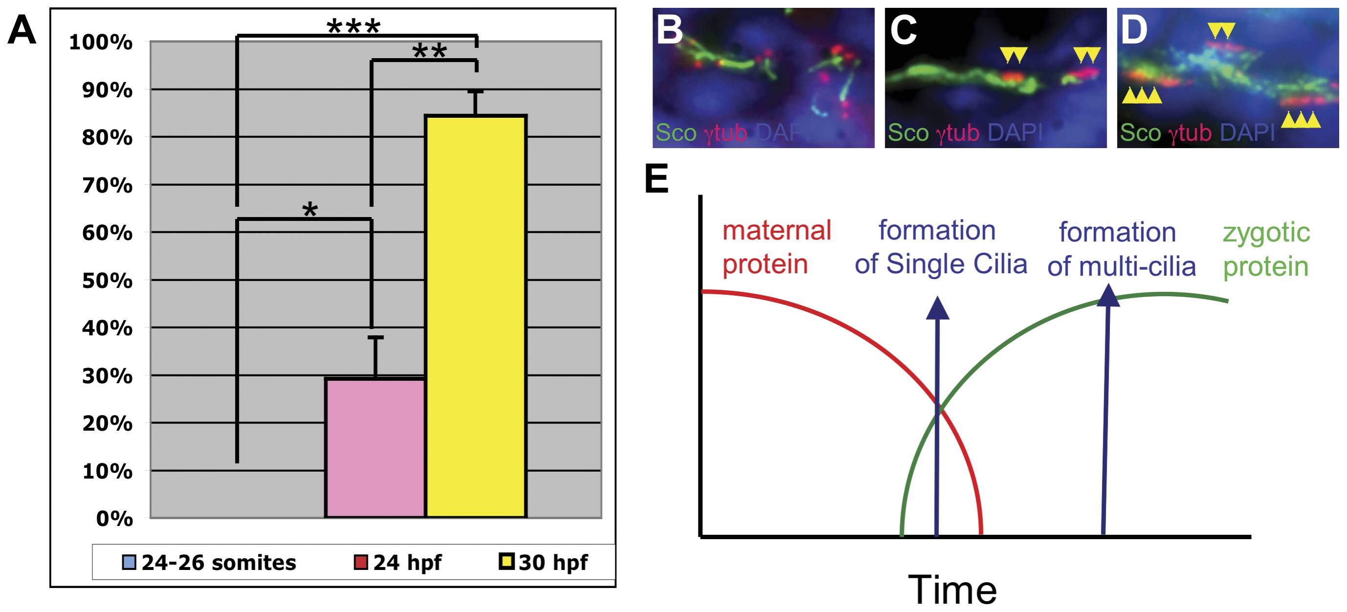
Fig. 5 Single cilia form earlier in development than multi-cilia in the pronephric duct.
(A) Graphical representation of multi-cilia observed in embryos at 24 somite, 24 hpf, and 30 hpf. Single cilia were observed at all time points analyzed. Bars represent percentage of embryos that developed multi cilia in the pronephric ducts. Each bar represents data from three independent experiments with at least 8 embryos each. (B–D) Representative images of cilia at 24-somite (B), 24 hpf (C) and 30 hpf (D). Yellow arrowheads point to basal bodies of multi-cilia. (E) A model of how different developmental timing of single cilia and multi-cilia could contribute to the pronephric cilia phenotypes observed in hi3959A mutants.