
IMAGE
Fig. 4
- ID
- ZDB-IMAGE-111213-12
- Antibodies
- Publication
- Li et al., 2011 - Qilin is essential for cilia assembly and normal kidney development in zebrafish
- All Figures
- Figures for Li et al., 2011
Image
|
Figure Caption
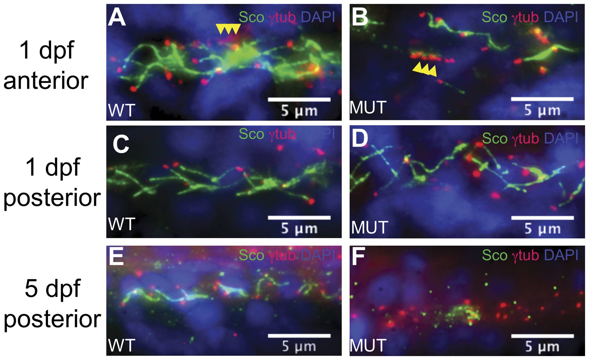
Fig. 4 Pronephric cilia in qilin hi3959A mutants are defective.
(A–D) Epifluorescent projections showing the pronephric cilia in a wild-type sibling (A, C) and a mutant (B, D) at the 1 dpf; in both anterior (A, B) and posterior (C,D) portions of the duct. Yellow arrowheads in A and B point to rows of basal bodies in MCCs. (E, F) Epifluorescent projections showing the pronephric cilia in a wild-type embryo (E) and a mutant (F) at 5 dpf in the posterior portion of the duct. All embryos were stained with anti-γ-tubulin (red), anti-Sco (green), and DAPI (blue).
Figure Data
Acknowledgments
This image is the copyrighted work of the attributed author or publisher, and
ZFIN has permission only to display this image to its users.
Additional permissions should be obtained from the applicable author or publisher of the image.
Full text @ PLoS One