
Fig. 3
- ID
- ZDB-IMAGE-111207-20
- Publication
- Ebert et al., 2005 - Calcium extrusion is critical for cardiac morphogenesis and rhythm in embryonic zebrafish hearts
- All Figures
- Figures for Ebert et al., 2005
|
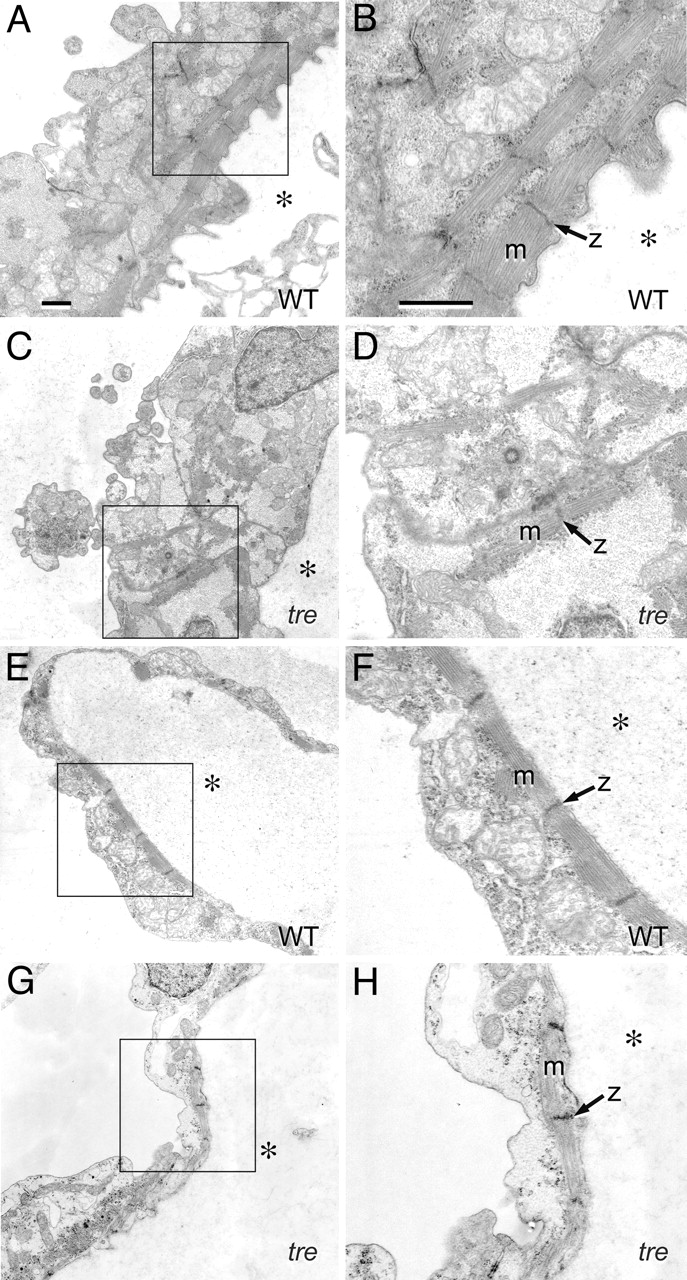
Fig. 3
Mutation of NCX1h is associated with myofibrillar defects in the ventricle. Transmission electron micrographs of embryonic hearts at 48 hpf. (A–D) Ventricle. In the wild-type ventricle (A), myofibrillar arrays are present along the interior aspect of the chamber and (B) bundled into consecutive units. In the tre ventricle (C), sarcomeres are scarce, randomly arrayed, and form few consecutive units. (D) Myofibrillar bundles are thin and lack distinct Z lines. (E–H) Atrium. In the atrium, both wild-type (E and F) and tre mutants (G and H) assemble consecutive sarcomeric units on the interior aspect of the chamber, with no substantial defects in tre mutants. However, the atrial cytoplasmic space in tre mutants (H) is electron-lucent, potentially indicative of decreased glycogen accumulation. (Bars, 1 μm.) B, D, F, and H are magnifications of boxed regions of A, C, E, and G, respectively. Magnification in A applies to C, E, and G; magnification in B applies to D, F, and H. * denotes the interior of the heart chamber.