
Fig. 2
- ID
- ZDB-IMAGE-111207-2
- Genes
- Antibodies
- Publication
- Jurynec et al., 2008 - Selenoprotein N is required for ryanodine receptor calcium release channel activity in human and zebrafish muscle
- All Figures
- Figures for Jurynec et al., 2008
|
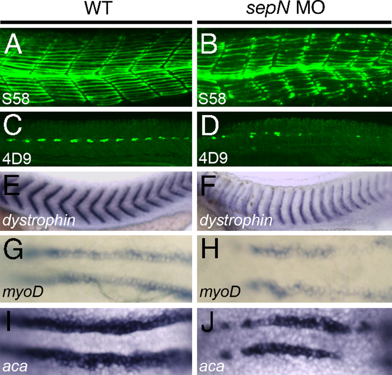
Fig. 2
Slow muscle fiber formation is reduced in sepN morphants. (A–F) Somitic muscle in midtrunk region of 24-hpf WT and sepN morphant embryos. (A and B) S58+ slow muscle fibers and (C and D) 4D9+ slow muscle pioneer fiber nuclei are present in reduced numbers in sepN morphants. Staining for dystrophin expression (E and F) reveals “u-shaped” somites in sepN morphant embryos. (G–J) Expression of myogenic lineage genes in adaxial cells of three-somite stage embryos. myoD (G and H) and α-cardiac actin (aca; I and J) are expressed discontinuously and at reduced levels in the adaxial cell population of sepN morphants. (A–F) Lateral views, rostral left. (G–J) Dorsal views, rostral left.