
Fig. 2
- ID
- ZDB-IMAGE-111129-7
- Publication
- Alexander et al., 2011 - Combinatorial roles for BMPs and Endothelin 1 in patterning the dorsal-ventral axis of the craniofacial skeleton
- All Figures
- Figures for Alexander et al., 2011
|
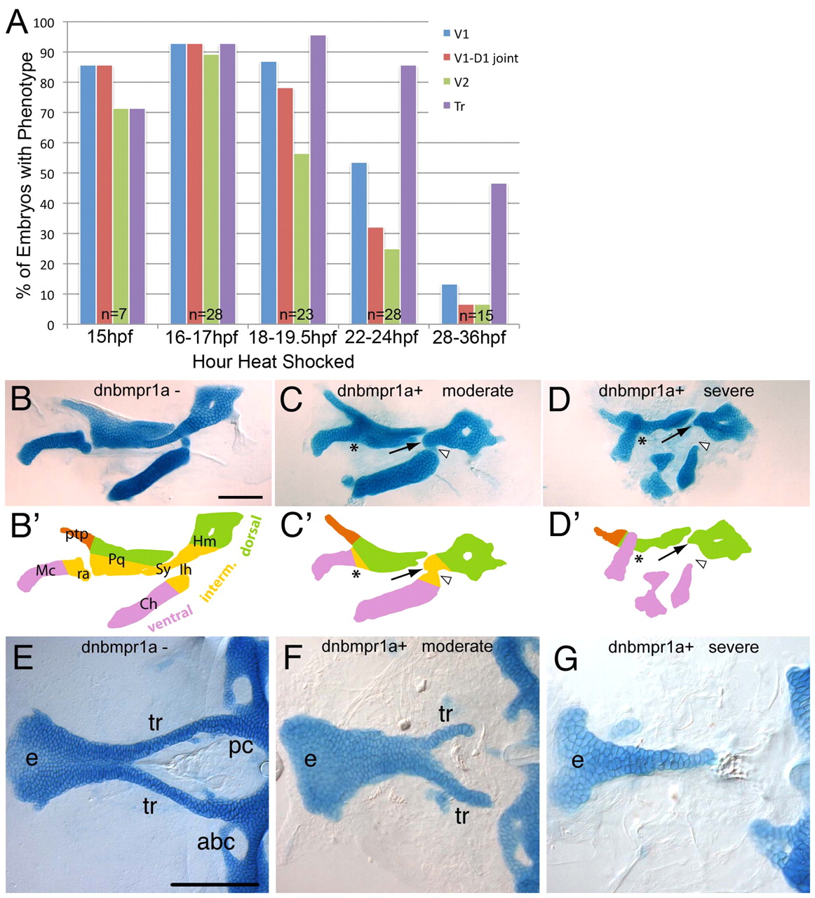
Fig. 2
Requirements for BMP signaling in pharyngeal cartilage and palate development. (A) Histogram quantifying the frequency of defects in different skeletal elements caused by heat shocking Tg(hs:dnBmpra1-GFP) embryos at different stages between 15 and 36 hpf. (B-G) Alcian Blue stained cartilages of 5 dpf larvae, dissected and flat-mounted, anterior towards the left. (B-D) Pharyngeal cartilages of control (B), moderate (C) and severely affected (D) Tg(hs:dnBmpra1-GFP) transgenics, heat shocked at 16-18 hpf, lateral views. (B2-D2) Diagrams of cartilage elements in arch 1 (mandibular, Mc and Pq) and arch 2 (hyoid, Ch, Ih and Hm) corresponding to cartilages in B-D. Lateral views, anterior towards the left. Colors indicate dorsal (green), intermediate (yellow) and ventral (pink) elements. Arrows indicate dorsal arch 2 elements; asterisks indicate fused joints in arch 1; arrowheads indicate joints in arch 2. (E-G) Neurocranial cartilages of the palate, ventral view: control (E), moderate (F) and severe (G). abc, anterior basicranial commissure; Ch, ceratohyal; e, ethmoid plate; Hm, hyomandibular; Ih, interhyal; Mc, Meckels cartilage; pc, parachordals; Pq, palatoquadrate; ptp, pterygoid process; ra, retroarticular process of Meckel’s cartilage; Sy, symplectic; tr, trabeculae. Scale bars: 100 μm.