
|
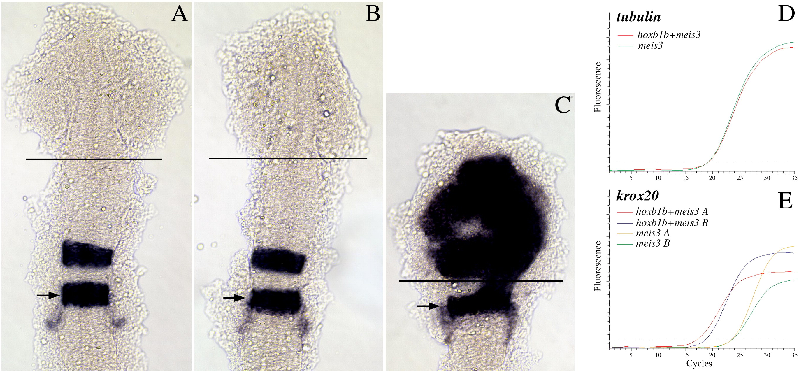
Fig. 1
An ectopic expression/dissection strategy to identify hoxb1b-regulated genes. (A–C) Uninjected (A), meis3 + βgal mRNA-injected (B) or meis3 + hoxb1b mRNA-injected (C) embryos were analyzed by in situ hybridization for expression of krox20. The position of r5 is indicated by arrows and the site where the anterior embryo was dissected is indicated by black lines in A–C. (D–E) Quantitative RT-PCR was used to assay expression of tubulin (D) and krox20 (E) in meis3-injected and meis3 + hoxb1b-injected embryos. Two independent injections are shown in E. Embryos in A–C were flat mounted at 14hpf and are shown in dorsal view with anterior to the top.
Reprinted from Developmental Biology, 358(2), Choe, S.K., Zhang, X., Hirsch, N., Straubhaar, J., and Sagerström, C.G., A screen for hoxb1-regulated genes identifies ppp1r14al as a regulator of the rhombomere 4 Fgf-signaling center, 356-67, Copyright (2011) with permission from Elsevier. Full text @ Dev. Biol.