
Image
|
Figure Caption
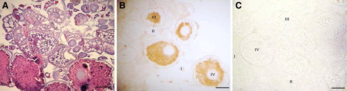
Fig. 5
Protein localization and distribution of the zebrafish Pumilio-2 in the ovary as detected by immunohistochemistry. a H&E staining, b DAB immunostained with the Pumillo-2 polyclonal antibody. c DAB immunostained with pre-immunization serum. I: stage I oocyte; II: stage II oocyte; III: stage III oocyte; IV: stage IV oocyte. N: nucleus. Scale bars: 0.3 mm in all panels
Figure Data
Acknowledgments
This image is the copyrighted work of the attributed author or publisher, and
ZFIN has permission only to display this image to its users.
Additional permissions should be obtained from the applicable author or publisher of the image.
Full text @ Mol. Biol. Rep.