
Image
|
Figure Caption
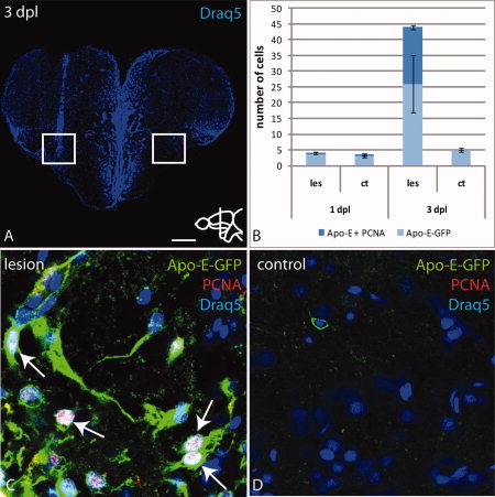
Fig. 8
Apo-E-GFP positive microglia cells proliferate in the parenchyma in response to wounding. A: Overview of a telencephalic cross-section through the lesioned region stained with Draq5. White box marks the ventral part of the lesion. B: Quantification of microglia cells labeled with Apo-E-GFP or together with PCNA. A subset of Apo-E-GFP microglia is proliferation-competent at 3 dpl. C,D: High magnification of the boxed region in A. Overlay of Apo-E-GFP, PCNA and Draq5 (C). Colocalization of PCNA and Apo-E-GFP shows that microglia proliferate near the lesion (arrows C). Scale bar = 100 μm in A; 7.5 μm in B–E.
Acknowledgments
This image is the copyrighted work of the attributed author or publisher, and
ZFIN has permission only to display this image to its users.
Additional permissions should be obtained from the applicable author or publisher of the image.
Full text @ Dev. Dyn.