
|
Fig. 7
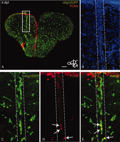
Stab injury leads to clustering of olig2:EGFP cells at the site of injury with a moderate proliferative response. A: Overview of a telencephalic cross-section through the injured region stained with anti-GFP and anti-PCNA antibodies at 6 dpl. The white box indicates olig2:EGFP cells clustering around the lesion. B–E: High magnification view of boxed region in A as maximum projection of a confocal Z-stack. The lesioned area is indicated by dashed lines. Draq5 staining in B shows the lesion filled with blood cells. Pictures in C–E show clustering of olig2:EGFP cells around the lesion (C, E) and a subpopulation of cells that is positive for proliferation nuclear antigen (PCNA) (arrows in D,E). Scale bar = 100 μm in A; 20 μm in B–E.