
|
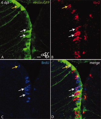
Fig. 6
At 6 dpl tbr2 positive cells are located intercalated between nestin:GFP radial glial cells and originated partly from progenitors that were in S-phase at 3 dpl. Bromodeoxyuridine (BrdU) was injected at 3 dpl followed by a chase until 6 dpl. A: Expression of nestin:GFP at 6 dpl. Note the up-regulation of nestin:GFP in the left hemisphere. B: Expression of tbr2 mRNA (arrows). C: BrdU incorporation (white arrows). D: Overlay of A–C. White arrows indicate tbr2 expressing cells positive for BrdU. These cells are intercalated between nestin:GFP positive cells. Yellow arrow shows a nestin:GFP cell also positive for tbr2 and BrdU. Scale bar = 10 μm.